. 2002 Jan 22;99(2):631-6.
doi: 10.1073/pnas.022531599.

Herpes simplex virus 1-infected cell protein 0 contains two E3 ubiquitin ligase sites specific for different E2 ubiquitin-conjugating enzymes

Affiliations

Herpes simplex virus 1-infected cell protein 0 contains two E3 ubiquitin ligase sites specific for different E2 ubiquitin-conjugating enzymes

Ryan Hagglund et al. Proc Natl Acad Sci U S A. .

Abstract

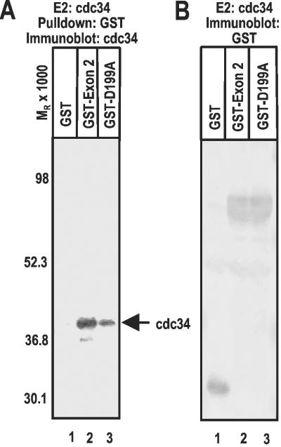
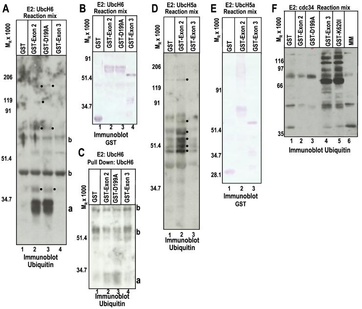
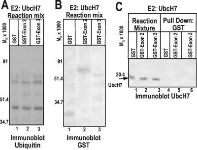
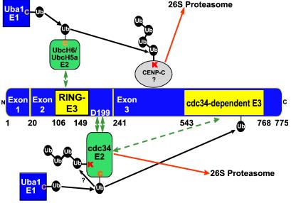
Infected cell protein 0 (ICP0) of herpes simplex virus 1, a multifunctional ring finger protein, enhances the expression of genes introduced into cells by infection or transfection, interacts with numerous cellular and viral proteins, and is associated with the degradation of several cellular proteins. Sequences encoded by exon 2 of ICP0 (residues 20-241) bind the UbcH3 (cdc34) ubiquitin-conjugating enzyme, and its carboxy terminus expresses a ubiquitin ligase activity demonstrable by polyubiquitylation of cdc34 in vitro. We report that: (i) The physical interaction of cdc34 and ICP0 leads to its degradation. Thus, substitution of ICP0 aspartate 199 with alanine attenuates the degradation of cdc34 and its binding to the ICP0 ring finger domain. (ii) Substitution of residue 620 reported to abolish the interaction with a ubiquitin-specific protease has no effect on the function of ubiquitin ligase. (iii) ICP0 contains an additional distinct E3 ligase activity specific for the UbcH5a- and UbcH6 E2-conjugating enzymes mapping to the ring finger domain. This is, to our knowledge, the first identification of a viral protein with at least two physically separated E3 ligase activities with different E2 specificities. The results suggest that each activity may target different proteins.


Figures

Figure 1
Figure 1
ICP0 mediates proteasome-dependent degradation of the E2 cdc34 in HSV-1-infected cells. (A) pHF infected with 10 plaque-forming units of HSV-1(F) (lanes 3 and 4) or R7914 (lanes 5 and 6) per cell were mock-treated or treated with MG132 for 1 h before harvest at 5 h after infection. Cells were solubilized, and lysates containing 100 μg of total protein were subjected to electrophoresis in denaturing polyacrylamide gels and reacted with a mouse monoclonal antibody against cdc34. (B) pHF were infected with HSV-1 and treated with MG132 as in A. The harvested cells were processed as above but probed with anti-ICP27 mouse monoclonal antibody. The bands were scanned and quantified as described in Materials and Methods.
Figure 2
Figure 2
Characterization of the interaction between cdc34 and sequences encoded by ICP0 exon 2. In vitro ubiquitylation reactions were performed as previously described (17). (A) GST (lane 1) or the indicated GST fusion protein (lanes 2 and 3) was pulled down with glutathione Sepharose beads from reactions containing cdc34, electrophoretically separated in a denaturing polyacrylamide gel, and reacted with anti cdc34 rabbit polyclonal antibody. (B) In vitro ubiquitylation reactions were performed by using cdc34 as in A. The proteins separated as above were reacted with anti-GST antibody. The bands were scanned and quantified as described in Materials and Methods.
Figure 3
Figure 3
ICP0 sequences encoded by exon 2 express an E3 ligase function. (A) In vitro ubiquitylation reactions were performed as previously described (17). The mixtures containing GST or the indicated GST fusion protein, E1, the E2 UbcH6, biotinylated Ub, ATP, and an ATP regenerating system were allowed to react for 30 min, then were subjected to electrophoresis in denaturing polyacrylamide gels and reacted with streptavidin HRP to detect biotinylated Ub. The dots to the right of the bands indicate polyubiquitylated species, which are specifically present or augmented in reactions containing GST-exon 2, either wild-type or carrying the D199A substitution. a, monoubiquitylated UbcH6; b, proteins present in reactions or ubiquitylated species formed independently of E3 activity, which reacted with streptavidin–HRP. (B) The same as A, except that the electrophoretically proteins were reacted with anti-GST antibody. The data serve as a loading control. (C) [His6]-UbcH6 was pulled down with talon resin from reaction mixtures containing UbcH6, GST, or the indicated GST fusion protein to determine whether the E2 was being ubiquitylated in the reactions (17), separated as above, and probed with streptavidin–HRP conjugate. a, monoubiquitylated UbcH6. Note the absence of polyubiquitylated UbcH6. (D) In vitro ubation reactions were done as in A but with E2 UbcH5a. The reaction mixture was probed for ubiquitylated species with streptavidin–HRP conjugate. The dots to the right of the bands indicate polyubiquitylated species, which are present or augmented in reactions containing GST-exon 2. (E) The electrophoretically separated reaction mix was reacted with anti-GST antibody as a loading control. (F) The experiment was set up as above but with the E2 cdc34. The objective was to test whether a mutation reported to abolish interaction with USP7 (36) affects the polyubiquitylation of cdc34 by ICP0 sequences encoded by exon 3. The reaction mixtures were separated on denaturing polyacrylamide gels and reacted with streptavidin–HRP conjugate to detect Ub. Note the presence of polyubiquitylated species in lanes 4 and 5.
Figure 4
Figure 4
ICP0 does not functionally or physically interact with the E2 enzyme UbcH7. (A) In vitro ubiquitylation reactions containing E2 UbcH7 were performed as in Fig. 3, then separated by SDS/PAGE and probed with streptavidin–HRP conjugate. (B) In vitro ubiquitylation reactions were performed and electrophoretically separated as in A and reacted with antibody directed against GST. (C) GST or GST fusion protein was pulled down with glutathione Sepharose beads from reaction mixtures containing UbcH7, separated as above, and probed with anti-UbcH7 mouse monoclonal antibody (lanes 4–6). Total reaction mixtures were also electrophoretically separated and reacted with mouse monoclonal antibody directed against UbcH7 (lanes 1–3).
Figure 5
Figure 5
Model for multiple specific E3 Ub ligase functions encoded by ICP0. The E3 Ub ligase function encoded by ICP0 exon 2 promotes Ub–protein ligation in conjunction with the E2 enzymes UbcH6 and UbcH5a. As ICP0 exon 2 contains the RING finger domain (amino acids 106–149), it is likely that this domain encodes a classical RING finger unimolecular E3 ligase (14, 20, 21). The accumulation of conjugated Ub and the degradation of cellular proteins by ICP0 require the RING finger (, –22, 24, 25), suggesting it is required for the E3 function involved in these processes. UbcH6 is presumed to transiently or weakly interact with the RING finger domain or other sequences within exon 2. ICP0 exon 3 encodes an unconventional RING finger-independent E3 ligase that functions in conjunction with the E2 cdc34. cdc34 interacts with ICP0 exon 2, and aspartate 199 is required for optimal interaction. The model proposes that the interaction tethers cdc34 to exon 3, which promotes the autoubiquitylation and degradation of cdc34. It is also possible that the cdc34-dependent E3 activity in exon 3 may act on additional substrates, although the degradation of cdc34 mediated by ICP0 would render such activity inefficient. This may provide the reason the virus evolved two discrete E3 activities in ICP0 to ensure the efficient degradation of cellular proteins. cdc34 is presumed to transiently or weakly interact with ICP0 exon 3. Transient or weak interactions are indicated by a double arrow. Strong interactions are indicated by two double green arrows. Numbers indicate the residue position in ICP0.

Similar articles

Cited by

References

    1. Roizman B, Knipe D M. In: Fields Virology. 4th Ed. Knipe D M, Howley P, Griffin D E, Lamb R A, Martin M A, Roizman B, Straus S E, editors. New York: Lippincott–Williams & Wilkins; 2001. pp. 2399–2459.
    1. Sacks W R, Schaffer P A. J Virol. 1987;61:829–839. - PMC - PubMed
    1. Stow N D, Stow E C. J Gen Virol. 1986;67:2571–2585. - PubMed
    1. Kawaguchi Y, Tanaka M, Yokoymama A, Matsuda G, Kato K, Kagawa H, Hirai K, Roizman B. Proc Natl Acad Sci USA. 2001;98:1877–1882. . (First Published February 6, 2001; 10.1073/pnas.041592598) - PMC - PubMed
    1. Kawaguchi Y, Bruni R, Roizman B. J Virol. 1997;71:1019–1024. - PMC - PubMed

Publication types

MeSH terms

LinkOut - more resources