. 2002 Jan 22;99(2):745-50.
doi: 10.1073/pnas.012585199.

Budding yeast Dsk2p is a polyubiquitin-binding protein that can interact with the proteasome

Affiliations

Budding yeast Dsk2p is a polyubiquitin-binding protein that can interact with the proteasome

Minoru Funakoshi et al. Proc Natl Acad Sci U S A. .

Abstract

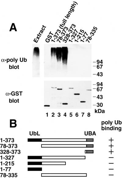
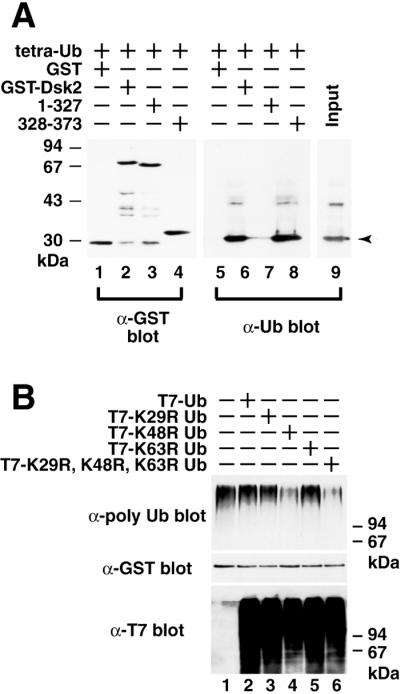
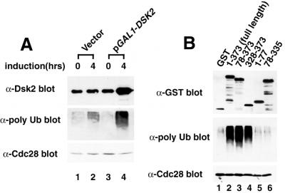
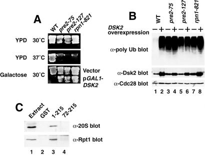
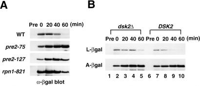
Dsk2p from Saccharomyces cerevisiae belongs to the class of proteins that contain a ubiquitin-like (UbL) domain at the N terminus together with a ubiquitin-associated (UBA) domain at the C terminus. We show here that the C-terminal UBA domain of Dsk2p binds to K48-linked polyubiquitin chains, and the N-terminal UbL domain of Dsk2p interacts with the proteasome. Overexpression of Dsk2p caused the accumulation of large amounts of polyubiquitin, and extragenic suppressors of the Dsk2p-mediated lethality proved to be temperature-sensitive mutations in two proteasome subunits, rpn1 and pre2. K48-linked ubiquitin-dependent degradation was impaired by disruption of the DSK2 gene. These results indicate that Dsk2p is K48-linked polyubiquitin-binding protein and also interacts with the proteasome. We discuss a possible role of adaptor function of Dsk2p via its UbL and UBA domains in the ubiquitin-proteasome pathway.


Figures

Figure 1
Figure 1
Dsk2p binds to polyubiquitin. (A) Interaction of Dsk2p with Ub was tested by two-hybrid assay. Among 1.5 × 10⁵ transformants screened, 17 UBI1∼3 and 4 DSK2 interacting clones were obtained. Interaction of DSK2 with UBI1∼3 was tested by histidine-prototrophic growth (Top) and β-gal (Middle). N-terminal truncated Dsk2p (residues 72–373) was used as a bait (Bottom), because overexpression of full-length Dsk2p is toxic to yeast (2). (B) Binding of GST-Dsk2p to Ub. GST-Dsk2p (lanes 2 and 4) and T7-Ub (lanes 3 and 4) were expressed in yeast. GST-Dsk2p was pulled down by glutathione beads, and GST-bound materials were immunoblotted by anti-GST (Upper) and anti-T7 antibodies (Lower). Lanes 1–4, expression; lanes 5–8, GST pull-down. (C) Binding of Ub to Dsk2p. His6-T7-Ub (lanes 2 and 4) and Dsk2p (lanes 1–4) were expressed in yeast. His6-T7-Ub was pulled down by Talon resin, and the bound materials (lanes 3 and 4) were immunoblotted either by anti-T7 (Upper) or anti-Dsk2 (Lower) antibody. An arrow indicates Dsk2p (see lane 4). (D) Dsk2p binds to polyubiquitin. GST (lane 2) or GST-Dsk2p (lane 3) purified from E. coli BL21 (DE3) was incubated with yeast extracts for 2 h. GST pull-down materials were immunoblotted with anti-polyubiquitin (lanes 2 and 3). Endogenous polyubiquitin in the extracts is shown in lane 1.
Figure 2
Figure 2
The C-terminal UBA domain of Dsk2p is required for its binding to polyubiquitin. (A) Binding assay of deletion mutants. Recombinant GST-Dsk2p deletions were incubated with yeast extract for 2 h, and GST-bound materials were immunoblotted either with the anti-polyubiquitin (Upper) or anti-GST (Lower) antibody. (B) A diagram of Dsk2p constructs. Binding abilities of the mutants are shown on the right.
Figure 3
Figure 3
The UBA domain of Dsk2p binds directly to K48-linked polyubiquitin chains. (A) Direct binding of Dsk2p to tetra-Ub. K48-linked tetra-Ub was incubated in vitro with full-length GST-Dsk2p (residues 1–373), C-terminal truncated GST-Dsk2p (residues 1–327), and C-terminal UBA (residues 328–373). GST pull-downs were immunoblotted with anti-GST (lanes 1–4) and anti-Ub (lanes 5–8) antibodies. The arrow in lane 9 indicates the position of tetra-Ub. (B) Effects of Ub lysine mutants on binding between Dsk2p and polyubiquitin. T7-tagged lysine mutants of Ub (K29R, K48R, K63R, and K29R-K48R-K63R) were expressed in yeast (Bottom), and the cell extracts were incubated with recombinant GST-Dsk2p. GST-bound materials were immunoblotted with anti-polyubiquitin (Top) and anti-GST (Middle) antibodies for GST-Dsk2p.
Figure 4
Figure 4
Dsk2p causes the accumulation of polyubiquitin chains. (A) Accumulation of polyubiquitin by overexpression of Dsk2p. Yeast cells transformed by pGAL1-DSK2 were incubated in raffinose synthetic medium (lanes 1 and 3), and Dsk2p expression was induced in 4 h by adding galactose to the medium (lanes 2 and 4). Proteins from the extracts were immunoblotted with anti-Dsk2p (Top), anti-polyubiquitin (Middle), and anti-Cdc28 (Bottom) antibodies. (B) The C-terminal UBA domain of Dsk2p is required for accumulation of polyubiquitin. GST-Dsk2p and its deletions (see Fig. 2) were galactose-induced in yeast, and proteins from the extracts were immunoblotted with anti-GST (Top), anti-polyubiquitin (Middle), and anti-Cdc28 (Bottom) antibodies.
Figure 5
Figure 5
Genetic and physical interactions between Dsk2p and the proteasomal subunits. (A) Identification of pre2 and rpn1. Suppressors were isolated as temperature-sensitive mutants that can suppress the Dsk2p-mediated growth arrest. The detailed procedure is described elsewhere (34). Mutation sites were determined by the gap-repair method as pre2-75, pre2-127, and rpn1-821. WT, wild type. (B) Polyubiquitin accumulates in pre2 and rpn1. DSK2 was overexpressed in pre2 and rpn1 at the permissive temperature (30°C), and cell extracts (lanes 2, 4, 6, and 8) were immunoblotted with anti-polyubiquitin (Top), anti-Dsk2p (Middle) and anti-Cdc28 (Bottom) antibodies. Lanes 1, 3, 5, and 7, no induction of Dsk2p. (C) Physical interaction of Dsk2p with the proteasome. GST-fused Dsk2p recombinant protein was incubated in the yeast cell extracts. GST pull-downs were immunoblotted with anti-20S (Upper) and anti-Rpt1 (Lower) antibodies. Lane 1, cell extracts; lane 2, GST control; lane 3, Dsk2p (residues 1–215); lane 4, N-terminal truncated Dsk2p (residues 72–215).
Figure 6
Figure 6
Inhibition of protein degradation by the proteasome mutations pre2 and rpn1 and by DSK2 disruption. (A) Inhibition of Leu-β-gal degradation in pre2 and rpn1. Ub-Leu-β-gal, a model substrate of N-end rule (originally provided by A. Varshavsky), was induced by galactose induction in the wild-type, pre2, and prn1 strains followed by chasing with the addition of glucose to the medium. After the induction was shut off, cell extracts (0.1 A600 equivalents) were taken at 20-min intervals and immunoblotted with anti-β-gal antibody (Promega). Pre; before induction; WT, wild type. (B) Degradation of K48-linked N-end rule substrate is inhibited by DSK2 disruption. N-end rule substrates, Ub-Leu-β-gal and Ub-Ala-β-gal, were induced by galactose-induction in dsk2Δ (lanes 1–5) and the wild-type strain (lanes 6–10). Degradation of Leu-β-gal and Ala-β-gal was followed by immunoblotting as described for A.

References

    1. Jentsch S, Pyrowlakis G. Trends Cell Biol. 2000;10:335–342. - PubMed
    1. Biggins S, Ivanovska I, Rose M D. J Cell Biol. 1996;133:1331–1346. - PMC - PubMed
    1. Schauber C, Chen L, Tongaonkar P, Vega I, Lambertson D, Potts W, Madura K. Nature (London) 1998;391:715–718. - PubMed
    1. Lambertson D, Chen L, Madura K. Genetics. 1999;153:69–79. - PMC - PubMed
    1. Ortolan T G, Tongaonkar P, Lambertson D, Chen L, Schauber C, Madura K. Nat Cell Biol. 2000;2:601–608. - PubMed

Publication types

MeSH terms