. 2002 Jan 22;99(2):751-6.
doi: 10.1073/pnas.022462899.

Actin cable dynamics in budding yeast

Affiliations

Actin cable dynamics in budding yeast

Hyeong-Cheol Yang et al. Proc Natl Acad Sci U S A. .

Abstract

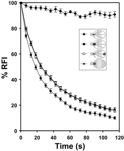
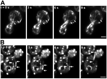
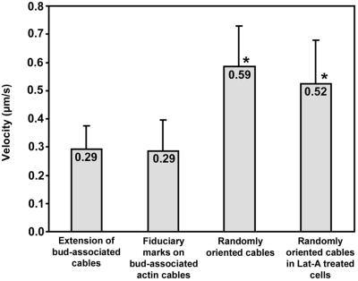
Actin cables, bundles of actin filaments that align along the long axis of budding yeast, are crucial for establishment of cell polarity. We fused green fluorescent protein (GFP) to actin binding protein 140 (Abp140p) and visualized actin cable dynamics in living yeast. We detected two populations of actin cables: (i) bud-associated cables, which extend from the bud along the mother-bud axis, and (ii) randomly oriented cables, which are relatively short. Time-lapse imaging of Abp140p-GFP revealed an apparent increase in the length of bud-associated actin cables. Analysis of movement of Abp140p-GFP fiduciary marks on bud-associated cables and fluorescence loss in photobleaching experiments revealed that this apparent elongation occurs by assembly of new material at the end of the cable within the bud and movement of the opposite end of the cable toward the tip of the mother cell distal to the bud. The rate of extension of the tip of an elongating actin cable is 0.29 +/- 0.08 microm/s. Latrunculin A (Lat-A) treatment completely blocked this process. We also observed movement of randomly oriented cables around the cortex of cells at a rate of 0.59 +/- 0.14 microm/s. Mild treatment with Lat-A did not affect the velocity of movement of randomly oriented cables. However, Lat-A treatment did increase the number of randomly oriented, motile cables per cell. Our observations suggest that establishment of bud-associated actin cables during the cell cycle is accomplished not by realignment of existing cables but by assembly of new cables within the bud or bud neck, followed by elongation.


Figures

Figure 1
Figure 1
(A) Colocalization of Abp140p–GFP (a) and the actin cytoskeleton (b). Cells (YCY006) expressing Abp140p–GFP were grown to midlog phase at room temperature (22–24°C) in lactate medium, fixed, and stained with rhodamine phalloidin to visualize F-actin structures. Fluorescence images of GFP and rhodamine were obtained at 0.3-μm z-intervals through the entire cell, deconvolved, and reconstructed. A two-dimensional projection of the three-dimensional reconstruction is shown. Arrows indicate regions along bud-associated actin cables that are brightly labeled with rhodamine phalloidin and Abp140p–GFP. Arrowhead shows a randomly oriented actin cable. Punctate structures in the mother cell and bud that show strong staining with rhodamine phalloidin and weak fluorescence with Abp140p–GFP are actin patches. (B) Fluorescence intensity of Abp140p–GFP and rhodamine phalloidin along an actin cable. The fluorescence images were obtained as in A, and fluorescence intensity was measured along the cable of Abp140p–GFP (a) and F-actin (b). The black and gray lines in c represent intensity of Abp140p–GFP and rhodamine phalloidin, respectively. The size of the area defining the actin cable for the measurement is 0.28 × 2.0 μm. (Bar, 1.5 μm.)
Figure 2
Figure 2
(A) Abp140p–GFP localization to cable-like structures in living yeast is sensitive to Lat-A. Cells were grown as in Fig. 1 and incubated with 200 μM Lat-A dissolved in DMSO (c and d), or an equivalent amount (0.5%) of DMSO (a and b) for 2 min. Abp140p–GFP fluorescence (a and c) and phase-contrast images (b and d) are shown. (Bar, 1.5 μm.) (B) Destabilization of actin cables correlates with loss of Abp140p–GFP in cable-like structures in living yeast. Abp140p–GFP fluorescence in the actin cable mutant (tpm1–2 tpmΔ2) and control strain (TPM1 tpmΔ2) at 25°C (a and c, respectively) and 1 min after a shift from 25°C to 34.5°C (b and d). (Bar, 1 μm.)
Figure 3
Figure 3
Dynamics of bud-associated actin cables. (A) Extension of an Abp140p–GFP cable. Cells were grown as in Fig. 1. Fluorescence images of Abp140p–GFP were collected at a single plane of focus at 1.5-s intervals. White arrows point to the position of the tip of the elongating cable at 0 s. Black arrows indicate a fiduciary mark on the elongating cable that moves toward the end of the mother cell distal to the bud. (Bar, 1 μm.) (B) Immobilization of bud-associated actin cables in Lat-A-treated cells. Lat-A was added to a final concentration of 35 μM to a midlog phase liquid culture (OD600 = 1.0), and an aliquot of the cell suspension was placed on a microscope slide. Time-lapse images of Abp140p–GFP-labeled actin cables in a budding cell were acquired as in A. The total time from addition of Lat-A to acquisition of the first image was 2 min. Arrows indicate the position of a fluorescence fiduciary mark at 0 s. (Bar, 1 μm.)
Figure 4
Figure 4
FLIP of Abp140p–GFP. Cells were prepared as in Fig. 3A. For Lat-A treatment, cells were exposed to 400 μM Lat-A for 5 min. Lat-A was solubilized in DMSO and added such that the final concentration of DMSO was 0.8%. The control cells were incubated with 0.8% DMSO for 5 min. The bleached area is shown as dark circles (Inset). Fluorescence of a region in the mother cell (shaded zone, Inset) was quantitated to obtain relative fluorescence intensity (defined in Materials and Methods). Fifteen cells with small buds were selected for each result. ⧫, unbleached; ●, bleached near bud neck; □, bleached near distal tip of mother cell; ▴, bleached near bud neck after Lat-A treatment. Error bars represent standard error.
Figure 5
Figure 5
Dynamics of randomly oriented actin cables. (A) Movement of randomly oriented actin cables in an untreated cell. Cells were grown as in Fig. 1. Time-lapse images of Abp140p–GFP-labeled actin cables in a budding cell were acquired as in Fig. 3A. The beginning and end of a motile cable are marked with white arrows. (B) Randomly oriented actin cable movement in Lat-A -treated cells. Lat-A treatment and image acquisitions were performed as in Fig. 3B. The ends of a cable moving toward the bud are marked with arrows and arrowheads. The ends of a cable moving away from the bud are marked with white arrows. (Bars, 1 μm.)
Figure 6
Figure 6
Velocities of Abp140p–GFP cable movement. Cells were grown as in Fig. 1. Extension of cables was measured as the change in position of the tips of bud-associated cables away from the bud as a function of time. For movement of fiduciary marks, we monitored the time-dependent change in position of the center of bright fluorescent regions along Abp140p–GFP-labeled actin cables. All movements associated with extension of bud-associated actin cables and movement of fiduciary marks on bud-associated cables were unidirectional and directed toward the tip of the mother cell distal to the bud. To measure the velocity of the movement of randomly oriented actin cables either in untreated cells or in cells treated with Lat-A (35 μM, 2 min), we followed movement of the leading tips of moving actin cables. All velocities are averages of 10–20 measurements. Cables that displayed weak fluorescence or loss of fluorescence caused by photobleaching were excluded from these measurements. Error bars represent SD. * indicate velocities that were significantly different from the velocity of extension of bud-associated actin cables (P < 0.01, t test).

References

    1. DeRosier D J, Tilney L G. J Cell Biol. 2000;148:1–6. - PMC - PubMed
    1. Adams A E M, Pringle J R. J Cell Biol. 1984;98:934–945. - PMC - PubMed
    1. Amberg D C. Mol Biol Cell. 1998;9:3259–3262. - PMC - PubMed
    1. Cheney R E, O'Shea M K, Heuser J E, Coelho M V, Wolenski J S, Espreafico E M, Forscher P, Larson R E, Mooseker M S. Cell. 1993;75:13–23. - PubMed
    1. Hill K L, Catlett N L, Weisman L S. J Cell Biol. 1996;135:1535–1549. - PMC - PubMed

Publication types

MeSH terms