. 2002 Feb;184(4):884-8.
doi: 10.1128/jb.184.4.884-888.2002.

Cell-surface-anchoring role of N-terminal surface layer homology domains of Clostridium cellulovorans EngE

Affiliations

Cell-surface-anchoring role of N-terminal surface layer homology domains of Clostridium cellulovorans EngE

Akihiko Kosugi et al. J Bacteriol. 2002 Feb.

Abstract

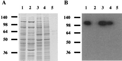
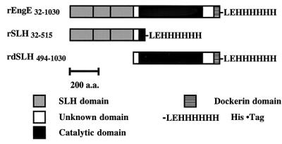
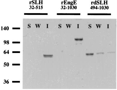
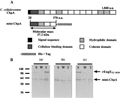
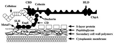
engE, coding for endoglucanase E, one of the three major subunits of the Clostridium cellulovorans cellulosome, has been cloned and sequenced (Y. Tamaru and R. H. Doi, J. Bacteriol. 181:3270-3276, 1999). The N-terminal-half region of EngE possesses three repeated surface layer homology (SLH) domains, which are homologous to those of some bacterial S-layer proteins. Also, the C-terminal-half region consists of a catalytic domain of glycosyl hydrolase family 5 and a duplicated sequence (dockerin) for binding EngE to scaffolding protein CbpA. Our hypothesis is that the SLH domains serve in the role of anchoring to the cell surface. This model was investigated by using recombinant EngEs (rEngE) with and without SLH domains that were synthesized in Escherichia coli and cell wall preparations from C. cellulovorans. When rEngE and SLH polypeptides of EngE were incubated with cell wall fragments prepared by sodium dodecyl sulfate treatment, these proteins bound strongly to the cell wall. However, rEngEs without SLH domains lost their ability to bind to cell walls. When rEngE was incubated with mini-CbpA, consisting of two cohesin domains, and cell wall fragments, the mini-CbpA was able to bind to the cell wall with rEngE. However, the binding of mini-CbpA was dramatically inhibited by addition of a chelating reagent, such as EDTA, which prevents cohesin-dockerin interactions. These results suggest not only that the SLH domains of EngE can bind to the cell surface but also that EngE plays an anchoring role for cellulosomes through the interaction of its dockerin domain with a CbpA cohesin.


Figures

FIG. 1.
FIG. 1.
Localization of EngE on C. cellulovorans. (A) Coomassie blue-stained SDS gel; (B) immunoblot with anti-EngE antiserum on each cell wall fraction. Lanes 1, whole cells (fraction F1); lanes 2, cell extract (fraction F2); lanes 3, crude cell walls (fraction F3); lanes 4, cell wall proteins (fraction F4); lanes 5, SDS-extracted cell wall fragments (fraction F5). Molecular size markers (in kilodaltons) are shown at the left. Four and 10 μl of each fraction were used for immunoblot analysis and SDS-PAGE, respectively.
FIG.2.
FIG.2.
Schematic representation of the domain structure of rEngE. The three rEngEs (rEngE32-1030, rSLH32-515, and rdSLH494-1030) were expressed by the pET system in E. coli BL21. Plasmid construction is described in Materials and Methods. These recombinant proteins were purified with a Ni affinity column. a.a., amino acids.
FIG. 3.
FIG. 3.
Binding of SLH domains of EngE to C. cellulovorans cell wall fragments. Each purified rEngE was incubated with fraction F5. Any insoluble material was precipitated and washed as described in Materials and Methods. Lanes S, soluble fraction; lanes W, wash fraction; lanes I, insoluble fraction. Molecular size markers (in kilodaltons) are shown at the left.
FIG. 4.
FIG. 4.
(A) Schematic representation of C. cellulovorans CbpA and mini-CbpA. a.a., amino acids. (B) Binding of mini-CbpA to the cell surface by SLH domains of EngE. Purified rEngE32-1030 and mini-CbpA were incubated with fraction F5 and analyzed by SDS-PAGE. (a) Reaction of rEngE32-1030 and mini-CbpA; (b) reaction of mini-CbpA only; (c) reaction of rEngE32-1030 and mini-CbpA plus 20 mM EDTA. Any insoluble material was precipitated and washed as described in Materials and Methods. Lanes S, soluble fraction; lanes W, wash fraction; lanes I, insoluble fraction. Molecular size markers (in kilodaltons) are shown at the left.
FIG. 5.
FIG. 5.
Model showing attachment of EngE to the CbpA of C. cellulovorans and the cell surface. The three repeated SLH domains of the N terminus of EngE integrate into the cell wall layer containing peptidoglycan, while the C terminus of EngE is bound to CbpA through its dockerin domain. The question mark indicates that some interactions between SLH domains of EngE and cell wall containing peptidoglycan may be bridged by divalent ions but the data are not conclusive. CBD, cellulose binding domain; CD, catalytic domain.

References

    1. Bayer, E. A., L. J. W. Shimon, Y. Shoham, and R. Lamed. 1998. Cellulosome--structure and ultrastructure. J. Struct. Biol. 124:221-234. - PubMed
    1. Bingle, W., J. F. Nomellini, and J. Smit. 1997. Linker mutagenesis of the Caulobacter crescentus S-layer protein: toward a definition of an N-terminal anchoring region and a C-terminal secretion signal and the potential for heterologous protein secretion. J. Bacteriol. 179:601-611. - PMC - PubMed
    1. Brechtel, E., and H. Bahl. 1999. In Thermoanaerobacterium thermosulfurigenes EM1 S-layer homology domains do not attach to peptidoglycan. J. Bacteriol. 181:5017-5023. - PMC - PubMed
    1. Chauvaux, S., M. Matuschek, and P. Béguin. 1999. Distinct affinity of binding sites for S-layer homologous domains in Clostridium thermocellum and Bacillus anthracis cell envelopes. J. Bacteriol. 181:2455-2458. - PMC - PubMed
    1. Doi, R. H., and Y. Tamaru. 2001. The Clostridium cellulovorans cellulosome: an enzyme complex with plant cell wall degrading activity. Chem. Rec. 1:24-32. - PubMed

Publication types

LinkOut - more resources