. 2002 Feb;184(4):1140-54.
doi: 10.1128/jb.184.4.1140-1154.2002.

Pseudomonas aeruginosa displays multiple phenotypes during development as a biofilm

Affiliations

Pseudomonas aeruginosa displays multiple phenotypes during development as a biofilm

Karin Sauer et al. J Bacteriol. 2002 Feb.

Abstract

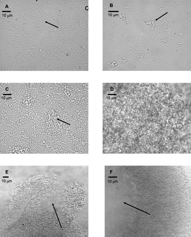
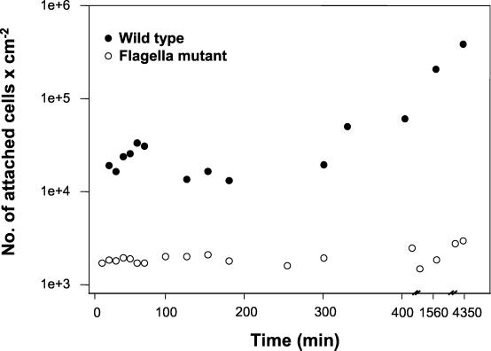
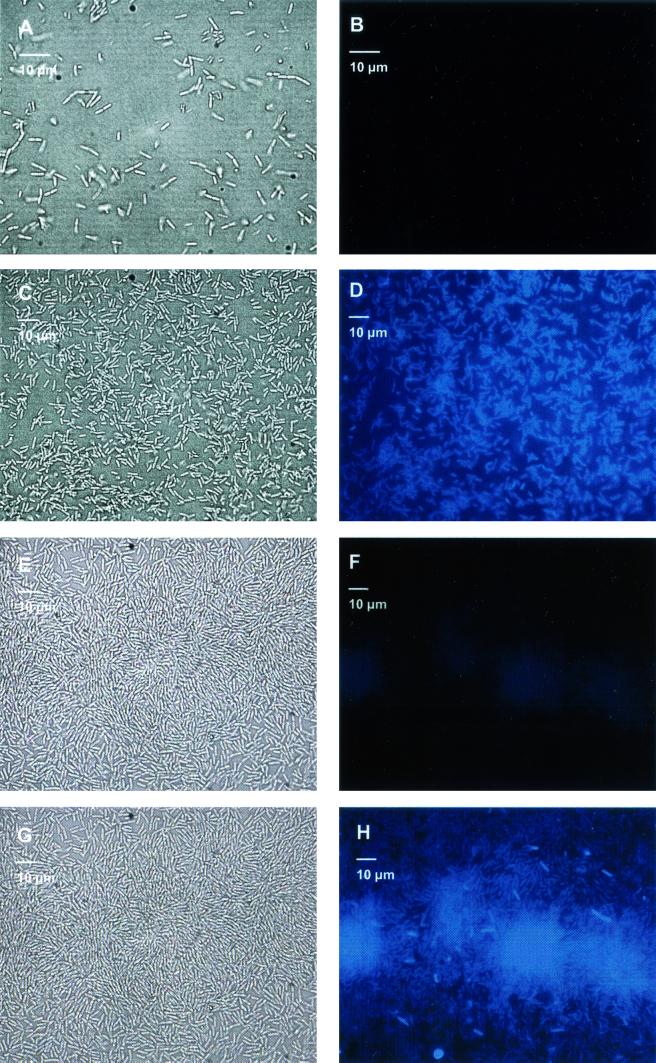
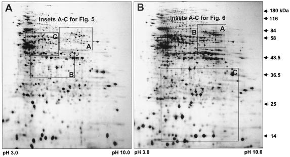
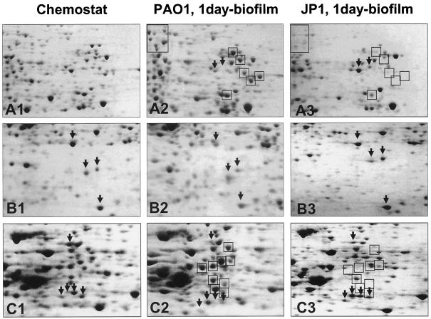
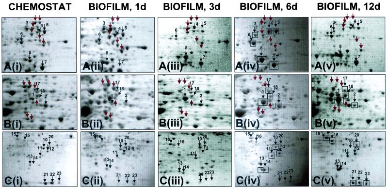
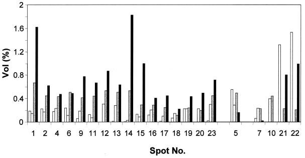
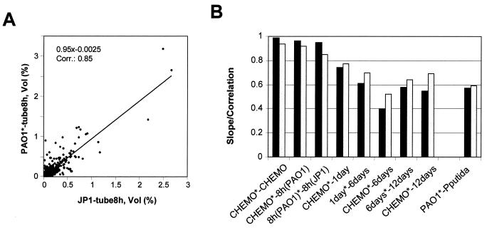
Complementary approaches were employed to characterize transitional episodes in Pseudomonas aeruginosa biofilm development using direct observation and whole-cell protein analysis. Microscopy and in situ reporter gene analysis were used to directly observe changes in biofilm physiology and to act as signposts to standardize protein collection for two-dimensional electrophoretic analysis and protein identification in chemostat and continuous-culture biofilm-grown populations. Using these approaches, we characterized five stages of biofilm development: (i) reversible attachment, (ii) irreversible attachment, (iii) maturation-1, (iv) maturation-2, and (v) dispersion. Biofilm cells were shown to change regulation of motility, alginate production, and quorum sensing during the process of development. The average difference in detectable protein regulation between each of the five stages of development was 35% (approximately 525 proteins). When planktonic cells were compared with maturation-2 stage biofilm cells, more than 800 proteins were shown to have a sixfold or greater change in expression level (over 50% of the proteome). This difference was higher than when planktonic P. aeruginosa were compared with planktonic cultures of Pseudomonas putida. Las quorum sensing was shown to play no role in early biofilm development but was important in later stages. Biofilm cells in the dispersion stage were more similar to planktonic bacteria than to maturation-2 stage bacteria. These results demonstrate that P. aeruginosa displays multiple phenotypes during biofilm development and that knowledge of stage-specific physiology may be important in detecting and controlling biofilm growth.


Figures

FIG. 1.
FIG. 1.
Transitional episodes in biofilm development by P. aeruginosa strain PAO1 examined by transmitted light microscopy. Each panel represents a distinct episode in biofilm development. (A) Reversible attachment. Initial event in biofilm development, bacteria are attached to substratum at cell pole (arrow). (B) irreversible attachment. Cells were cemented to the substratum and formed nascent cell clusters (arrow) with all cells in contact with the substratum. (C) Maturation-1. Cell clusters matured (arrow) and were several cells thick, embedded in the EPS matrix. (D) Maturation-2. Cell clusters reached maximum thickness, approximately 100 μm. (E and F) Dispersion. Cells evacuated interior portions of cell clusters (arrow), forming void spaces.
FIG. 2.
FIG. 2.
Effect of flagella on biofilm formation. Time course showing number of chemostat-grown cells attached to a glass substratum over a period of 3 days under conditions of continuous flow. •, P. aeruginosa strain PA14 (wild-type) cells attached at a constant rate, rapidly increasing the number of cells at the substratum. ○, mutants (P. aeruginosa strain PA257) defective in flagellum synthesis attached poorly and showed no significant increase in surface cell numbers over a period of 3 days.
FIG. 3.
FIG. 3.
Onset of quorum-sensing regulons in developing biofilm. (A) Transmitted light micrograph of P. aeruginosa strain PAO230 during the reversible attachment phase of biofilm development; (B) the same image under UV illumination demonstrated no activation of the Las regulon. (C) Transmitted light micrograph of P. aeruginosa strain PAO230 during the irreversible attachment phase of biofilm development; (D) the same image under UV illumination demonstrated activation of the Las regulon. (E) Transmitted light micrograph of P. aeruginosa strain PAO220 during the irreversible attachment phase of biofilm development; (F) the same image under UV illumination demonstrated no activation of the Rhl quorum-sensing regulon. (G) Transmitted light micrograph of P. aeruginosa strain PAO220 during the maturation-1 phase of biofilm development; (H) the same image under UV illumination demonstrated activation of the Rhl quorum-sensing regulon.
FIG. 4.
FIG. 4.
Two-dimensional images of crude protein extracts of P. aeruginosa grown as a 1-day biofilm (A) and as a 12-day biofilm (B). The crude protein extracts (200 μg) were separated on pH 3 to 10 nonlinear Immobiline Dry strips (Amersham Pharmacia, Piscataway, N.J.), followed by SDS-11% polyacrylamide gel electrophoresis. Gels were stained with silver nitrate (3). Outlined areas in panel A indicate zones of the two-dimensional gels that are represented in Fig. 5, and outlined areas in panel B indicate zones represented in Fig. 6.
FIG. 5.
FIG. 5.
Enlarged partial two-dimensional gels showing crude protein extract of P. aeruginosa strain PAO1 grown planktonically (A1 to C1), attached for 1 day (A2 to C2) as well as P. aeruginosa JP1 after 1 day of attachment time (A3 to C3). Boxed protein spots indicate proteins that are produced at higher levels in 1-day P. aeruginosa strain PAO1 biofilm cells than in 1-day strain PAO-JP1 biofilm cells; arrows pointing downward indicate proteins that were absent or only weakly expressed in 1-day P. aeruginosa strain PAO1 biofilm cells compared to strain PAO-JP1. The crude protein extracts (500 μg) were extracted and separated on pH 3 to 10 nonlinear Immobiline Dry strips, followed by SDS-11% PAGE. The gels were stained with Coomassie brilliant blue R350, Dashed-box protein spots were identified by mass spectrometry (see Fig. 6 and Table 2).
FIG. 6.
FIG. 6.
Enlarged partial two-dimensional gels showing crude protein extract of P. aeruginosa strain PAO1 grown planktonically in a chemostat [A(i) to C(i)] and for 1 day [A(ii) to C(ii)], 3 days [A(iii) to C(iii)], 6 days [A(iv) to C(iv)], and 12 days [A(v) to C(v)] in a biofilm. Sections A to C show an enlarged view of the two-dimensional images in Fig. 4. Proteins that were identified by MS analysis are indicated by boxes. The spot numbers correlate with the numbers given in Table 2. The gels were stained with Coomassie brilliant blue R-350.
FIG. 7.
FIG. 7.
Change in spot intensity and differential expression of selected proteins over the course of biofilm development. From left to right in each group of four, bars represent spot intensity obtained under planktonic growth conditions and after 1 day, 6 days, and 12 days of biofilm development. The spot numbers correlate with the numbers given in Table 2 and Fig. 6. The protein spots are organized following their protein expression pattern (upregulated, downregulated, and varied expression). Vol (%), relative volume (according to Melanie 3.0 software description), volume divided by the total volume over the whole image.
FIG. 8.
FIG. 8.
Representative scatter plot graph (A) and analysis of two-dimensional gel similarities by scatter plot (B). Solid bars, linear dependence; shaded bars, correlation; ∗, reference gel for the scatter plot analysis; CHEMO, chemostat-grown P. aeruginosa strain PAO1; 8 h, 1 day, 6 days, and 12 days, P. aeruginosa strain PAO1 biofilm after 8 h, 1 day, 6 days, and 12 days of attachment time, respectively; tube 8 h, biofilm after 8 h of attachment time; JP1, P. aeruginosa strain PAO-JP1; PAO1, chemostat-grown P. aeruginosa strain PAO1; P. putida (ATCC 39168), chemostat grown. Vol (%), relative volume (according to Melanie 3.0 software description), volume divided by the total volume over the whole image.

References

    1. Arai, H., T. Kodama, and Y. Igarashi. 1997. Cascade regulation of the two CRP/FNR-related transcriptional regulators (ANR and DNR) and the denitrification enzymes in Pseudomonas aeruginosa. Mol. Microbiol. 25:1141-1148. - PubMed
    1. Belas, R., M. Simon, and M. Silverman. 1986. Regulation of lateral flagella gene transcription in Vibrio parahaemolyticus. J. Bacteriol. 167:210-218. - PMC - PubMed
    1. Blum, H., H. Beier, and H. J. Gross. 1987. Improved silver staining of plant proteins, RNA and DNA in polyacrylamide gels. Electrophoresis 8:93-99.
    1. Boels, I. C., A. Ramos, M. Kleerebezem, and W. M. de Vos. 2001. Functional analysis of the Lactococcus lactis galU and galE genes and their impact on sugar nucleotide and exopolysaccharide biosynthesis. Appl. Environ. Microbiol. 67:3033-3040. - PMC - PubMed
    1. Bollinger, N., D. J. Hassett, B. H. Iglewski, J. W. Costerton, and T. R. McDermott. 2001. Gene expression in Pseudomonas aeruginosa: evidence of iron override effects on quorum sensing and biofilm-specific gene regulation. J. Bacteriol. 183:1990-1996. - PMC - PubMed

Publication types

MeSH terms