. 2002 Feb;184(4):1219-24.
doi: 10.1128/jb.184.4.1219-1224.2002.

Localization of the cortex lytic enzyme CwlJ in spores of Bacillus subtilis

Affiliations

Localization of the cortex lytic enzyme CwlJ in spores of Bacillus subtilis

Irina Bagyan et al. J Bacteriol. 2002 Feb.

Abstract

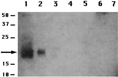
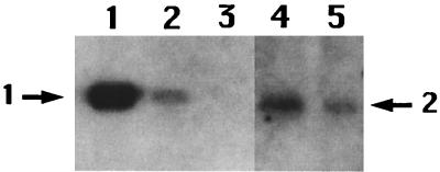
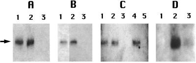
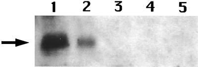
The enzyme CwlJ is involved in the depolymerization of cortex peptidoglycan during germination of spores of Bacillus subtilis. CwlJ with a C-terminal His tag was functional and was extracted from spores by procedures that remove spore coat proteins. However, this CwlJ was not extracted from disrupted spores by dilute buffer, high salt concentrations, Triton X-100, Ca(2+)-dipicolinic acid, dithiothreitol, or peptidoglycan digestion, disappeared during spore germination, and was not present in cotE spores in which the spore coat is aberrant. These findings indicate the following: (i) the reason decoated and cotE spores germinate poorly with dipicolinic acid is the absence of CwlJ from these spores; and (ii) CwlJ is located in the spore coat, presumably tightly associated with one or more other coat proteins.


Figures

FIG. 1.
FIG. 1.
Localization of CwlJ in spore fractions. Samples were prepared from spores of strains PS832 (wild-type) and IB650 (cwlJ-His-tag) and run on an SDS-polyacrylamide gel, proteins were transferred to an Immobilon-P membrane, and His-tagged proteins were detected as described in the text. The samples run in the various lanes are as follows: lanes 1 and 3, protein from cwlJ-His-tag or wild-type spores, respectively, extracted at an OD600 of 2.5 by the alkaline decoating procedure; lanes 2 and 4, insoluble fraction of disrupted cwlJ-His-tag or wild-type spores, respectively, at an OD600 of 2.5; lane 5, insoluble fraction from disrupted decoated cwlJ-His-tag spores at an OD600 of 2.5; lanes 6 and 7, soluble fractions of cwlJ-His-tag and wild-type spores, respectively, at an OD600 of 2.5. The numbers to the left of the figure denote the migration positions of molecular size markers in kDa; the arrow denotes the position of the CwlJ-His-tag protein. This figure, as well as all others, was processed in Adobe Photoshop, and all adjacent lanes are from a single SDS-PAGE experiment.
FIG. 2.
FIG. 2.
Level of CwlJ in spores. Various amounts of the P16-His-tag protein (22) or the insoluble fraction from cwlJ-His-tag spores were run on SDS-polyacrylamide gels, and His-tagged proteins were detected as described in the text. The samples run in the various lanes are the following: lanes 1, 2, and 3, 1 μg, 100 ng, and 10 ng, respectively, of the P16-His-tag protein, corresponding to 4 × 10¹³, 4 × 10¹², and 4 × 10¹¹ molecules, respectively; lanes 4 and 5, insoluble fractions of spores at OD600s of 5 and 2.5, respectively. The arrow labeled 1 denotes the migration position of the P16-His-tag protein; the arrow labeled 2 denotes the position of the CwlJ-His-tag protein. In this figure some intervening lanes on the original Western blot have been removed.
FIG. 3.
FIG. 3.
Lack of solubilization of CwlJ by cortex digestion, high-salt conditions, or Triton X-100, and disappearance of CwlJ upon spore germination. (A, B, and C) Spores of strain IB650 (cwlJ-His-tag) were disrupted, and the insoluble fraction was isolated and incubated with lysozyme (A), lysozyme and then 1 M NaCl (B), or 1% Triton X-100 (C) with or without prior lysozyme treatment as described in the text. After incubation, the samples were centrifuged, aliquots of the soluble and insoluble fraction were run on an SDS-polyacrylamide gel, and CwlJ was detected by Western blot analysis as described in the text. Note that in panel C insoluble protein was isolated and suspended in buffer containing 1% Triton X-100 prior to being mixed with sample buffer and boiled, since Triton X-100 interfered slightly with the Western blot analysis. (D) Spores of strain IB652 (cwlJ-His-tag ger3) were germinated with Ca2+-DPA and washed as described in the text, these spores as well as dormant spores were disrupted, and soluble and insoluble fractions were isolated. Aliquots equivalent to original dormant spores of various strains at an OD600 of 5 were loaded in each well, and the samples run on the various lanes are the following. (A) Lane 1, insoluble fraction prior to lysozyme treatment; lane 2, insoluble fraction after treatment with lysozyme; lane 3, protein solubilized by treatment of the insoluble fraction with lysozyme. (B) Lane 1, insoluble fraction after treatment with lysozyme; lane 2, insoluble fraction after treatment with lysozyme and then extraction with 1 M NaCl; lane 3, protein solubilized by extraction of the lysozyme-treated insoluble fraction with 1 M NaCl. (C) Lane 1, insoluble fraction prior to extraction with 1% Triton X-100; lane 2, insoluble fraction after extraction with Triton X-100; lane 3, protein solubilized from the insoluble fraction by Triton X-100; lane 4, insoluble fraction after digestion with lysozyme followed by extraction with Triton X-100; lane 5, protein solubilized by extraction of the lysozyme-treated insoluble fraction with Triton X-100. (D) Lane 1, soluble fraction from germinated spores; lane 2, insoluble fraction from dormant spores; lane 3, insoluble fraction from germinated spores. The arrow denotes the position of the 18-kDa CwlJ-His-tag protein. In panel B, some intervening lanes on the original Western blot have been removed.
FIG. 4.
FIG. 4.
Level of CwlJ in cotE spores. Spores of strains IB650 (cwlJ-His-tag) and IB673 (cotE cwlJ-His-tag) were disrupted, the soluble and insoluble fractions were isolated, aliquots were run on an SDS-polyacrylamide gel, and CwlJ-His-tag proteins were identified by Western blot analysis as described in the text. The samples run in the various lanes are the following: lanes 1 and 2, insoluble fractions of cwlJ-His-tag spores at OD600s of 5 and 2.5, respectively; lanes 3 and 4, insoluble fractions of cotE cwlJ-His-tag spores at OD600s of 5 and 10, respectively; lane 5, soluble fraction of cotE cwlJ-His-tag spores at an OD600 of 7.5. The arrow denotes the position of the 18-kDa CwlJ-His-tag protein.

References

    1. Anagnostopoulos, C., and J. Spizizen. 1961. Requirements for transformation in Bacillus subtilis. J. Bacteriol. 81:74-76. - PMC - PubMed
    1. Buchanan, C. E., and S. L. Neyman. 1986. Correlation of penicillin-binding protein composition with different functions of two membranes in Bacillus subtilis forespores. J. Bacteriol. 165:498-503. - PMC - PubMed
    1. Boland, F. M., A. Atrih, H. Chirakkal, S. J. Foster, and A. Moir. 2000. Complete spore-cortex hydrolysis during germination of Bacillus subtilis 168 requires SleB and YpeB. Microbiology 146:57-64. - PubMed
    1. Driks, A. 1999. The Bacillus subtilis spore coat. Microbiol. Mol. Biol. Rev. 63:1-20. - PMC - PubMed
    1. Driks, A., and P. Setlow. 1999. Morphogenesis and properties of the bacterial spore, p. 191-218. In Y. V. Brun and L. J. Shimkets (ed.), Prokaryotic development. American Society for Microbiology, Washington, D.C.

Publication types

MeSH terms

LinkOut - more resources