. 2002 Jan 21;156(2):241-8.
doi: 10.1083/jcb.200107135. Epub 2002 Jan 21.

The conserved Pkh-Ypk kinase cascade is required for endocytosis in yeast

Affiliations

The conserved Pkh-Ypk kinase cascade is required for endocytosis in yeast

Amy K A deHart et al. J Cell Biol. .

Abstract

Internalization of activated signaling receptors by endocytosis is one way cells downregulate extracellular signals. Like many signaling receptors, the yeast alpha-factor pheromone receptor is downregulated by hyperphosphorylation, ubiquitination, and subsequent internalization and degradation in the lysosome-like vacuole. In a screen to detect proteins involved in ubiquitin-dependent receptor internalization, we identified the sphingoid base-regulated serine-threonine kinase Ypk1. Ypk1 is a homologue of the mammalian serum- and glucocorticoid-induced kinase, SGK, which can substitute for Ypk1 function in yeast. The kinase activity of Ypk1 is required for receptor endocytosis because mutations in two residues important for its catalytic activity cause a severe defect in alpha-factor internalization. Ypk1 is required for both receptor-mediated and fluid-phase endocytosis, and is not necessary for receptor phosphorylation or ubiquitination. Ypk1 itself is phosphorylated by Pkh kinases, homologues of mammalian PDK1. The threonine in Ypk1 that is phosphorylated by Pkh1 is required for efficient endocytosis, and pkh mutant cells are defective in alpha-factor internalization and fluid-phase endocytosis. These observations demonstrate that Ypk1 acts downstream of the Pkh kinases to control endocytosis by phosphorylating components of the endocytic machinery.


Figures
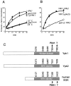
Figure 1.
Figure 1.
Ypk1 is required for α-factor internalization. (A and B) Internalization of 35S-α-factor was measured by the continuous presence protocol at 37°C (except where noted) after growth in YPUAD. ypk1 G490R cells are the same as udi5–1 cells. (A) YPK1 (LHY291, •); ypk1 G490R (LHY2543 ⋄); ypk1 G490R with pYPK1, a centromeric plasmid (LHY2712, ♦); ypk1 G490R (LHY2543, ○, at 24°C). (B) YPK1 YPK2 (LHY2632, •); ypk1Δ YPK2 (LHY2536, □); YPK1 ypk2Δ cells (LHY2633, ⋄). (C) Schematic diagrams of Ypk1, Ypk2 (68% identical to Ypk1), and human SGK (50% identical to Ypk1). Residues mutated in this study and their counterparts in Ypk1 homologues are shown. The percent identity of the kinase domains is shown in the gray box. Phosphorylation sites are indicated with an arrow with known kinases noted.
Figure 2.
Figure 2.
Ypk1 is required for fluid-phase endocytosis. (A and B) Lucifer yellow (LY) localization was assayed in ypk1 G490R (LHY2543), ypk1Δ (LHY2536), and wild-type cells from the same tetrad as each mutant (LHY2761 and LHY2537, respectively). Cells were grown to early logarithmic phase in rich medium at 24°C, shifted to 37°C for 15 min, and then allowed to internalize LY at 37°C for 30 or 60 min (A) Images were taken using DIC optics (top) and fluorescence optics (bottom). (B) The percent of cells that accumulated LY in their vacuoles was quantified for the 30- and 60-min time points by blind counting of each sample. The number of cells counted is indicated to the right of the corresponding bar.
Figure 3.
Figure 3.
The kinase activity of Ypk1 is required for receptor internalization, but not for receptor modifications. (A) Extracts of ypk1Δ cells expressing hemagglutinin (HA)-tagged wild-type Ypk1, Ypk1K376R, or Ypk1D488A were subjected to immunoblotting with α-HA antibodies. (B and C) Cells were grown and assayed for α-factor internalization as in Fig. 1. (B) The same cells used in A: ypk1Δ (LHY2536, •); YPK1 (LHY2563, ⋄); ypk1 K376R (LHY2564, ♦); ypk1 D488A (LHY2565, ○). (C) YPK1 cells expressing Ste2–378Stop (LHY825, •) or Ste2-Ub (LHY558, ♦); ypk1 G490R cells expressing Ste2–378Stop (LHY2684 ○) or Ste2-Ub (LHY2690 ⋄). (D) ste2Δ (LHY10), ypk1 G490R (LHY2543), or YPK1 (LHY1) cells were treated (+) or not (−) with 1 μM α-factor for 8 min at 37°C. Cell lysates were resolved by SDS-PAGE, transferred to nitrocellulose, and probed with α-Ste2 antibodies. Phosphorylated (P) and monoubiquitinated (Ub) species are denoted with arrows.
Figure 4.
Figure 4.
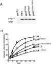
The conserved phosphorylation sites of Ypk1 are required for α-factor internalization. (A) Extracts of ypk1Δ cells expressing HA-tagged wild-type Ypk1, Ypk1T504A, Ypk1T662A, or Ypk1T504A,T662A . (B) The same cells used in A were grown and assayed as in Fig. 1: ypk1Δ (LHY2536, ▪); YPK1 (LHY2563, ♦); ypk1 T504A (LHY2568 X); ypk1 T662A (LHY2567, □); ypk1 T504A,T662A (LHY2569, ⊠).
Figure 5.
Figure 5.
Pkh kinases are required for receptor-mediated and fluid-phase endocytosis. (A) Growth of pkh1Δ pkh2Δ cells and pkh1Δ pkh2Δ leu2::PKH1 cells from the same tetrad (LHY2716 and LHY2714, respectively) or pkh ts (pkh1 D398G pkh2Δ) cells (LHY3030) and an isogenic pkh2Δ strain (LHY3032) on YPUAD at 24°C or 30°C or on YPUAD + 1.2 M sorbitol at 30°C after 4 d. The differences in strain background between the two pkh mutants are likely to account for the difference in suppression of the growth defect on sorbitol medium. (B) The same pkh1Δ pkh2Δ (♦) and pkh1Δ pkh2Δ leu2::PKH1 (⋄) strains as in (A) were grown overnight in YPUAD + 1.2M sorbitol. Internalization of 35S-α-factor was measured by the continuous presence protocol at 30°C in YPUAD + 1.2 M sorbitol. (C) LY localization was assayed for wild-type cells (LHY2762), pkh1Δ cells (LHY2759), pkh2Δ cells (LHY2760), pkh1Δ pkh2Δ cells (LHY2716), and end4Δ cells (LHY37). Cells were grown to early logarithmic phase in YPUAD + 1.2 M sorbitol at 24°C, shifted to 30°C for 15 min, and then incubated with LY at 30°C for 60 min. Images were taken using DIC optics (top) and fluorescence optics (bottom). The pkh1Δ pkh2Δ cells that are brightly stained throughout the whole cell are probably lysed cells. (D) pkh1Δ cells (LHY3031, •), pkh2Δ cells (LHY3032, ▪), and pkh ts cells (LHY3030, ♦) were grown overnight in YPUAD. Internalization of 35S-α-factor was measured by the continuous presence protocol at 30°C in YPUAD.

References

    1. Barbieri, M.A., A.D. Kohn, R.A. Roth, and P.D. Stahl. 1998. Protein kinase B/akt and Rab5 mediate Ras activation of endocytosis. J. Biol. Chem. 273:19367–19370. - PubMed
    1. Bauerfeind, R., K. Takei, and P. De Camilli. 1997. Amphiphysin I is associated with coated endocytic intermediates and undergoes stimulation-dependent dephosphorylation in nerve terminals. J. Biol. Chem. 272:30984–30992. - PubMed
    1. Casamayor, A., P.D. Torrance, T. Kobayashi, J. Thorner, and D.R. Alessi. 1999. Functional counterparts of mammalian protein kinases PDK1 and SGK in budding yeast. Curr. Biol. 9:186–197. - PubMed
    1. Chen, H., V.I. Slepnev, P.P. Di Fiore, and P. De Camilli. 1999. The interaction of epsin and Eps15 with the clathrin adaptor AP-2 is inhibited by mitotic phosphorylation and enhanced by stimulation-dependent dephosphorylation in nerve terminals. J. Biol. Chem. 274:3257–3260. - PubMed
    1. Chen, P., K.S. Lee, and D.E. Levin. 1993. A pair of putative protein kinase genes (YPK1 and YPK2) is required for cell growth in Saccharomyces cerevisiae. Mol. Gen. Genet. 236:443–447. - PubMed

Publication types

MeSH terms