. 2002 Jan 21;156(2):261-70.
doi: 10.1083/jcb.200110074. Epub 2002 Jan 21.

A new role for BiP: closing the aqueous translocon pore during protein integration into the ER membrane

Affiliations

A new role for BiP: closing the aqueous translocon pore during protein integration into the ER membrane

Nora G Haigh et al. J Cell Biol. .

Abstract

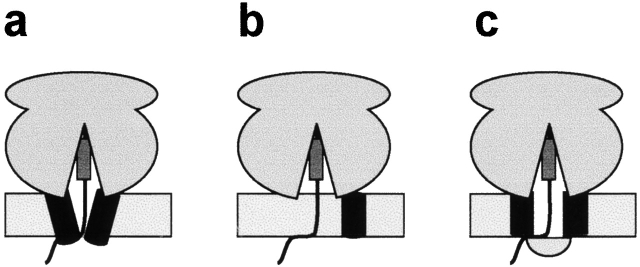
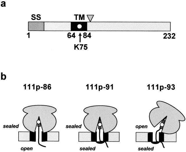
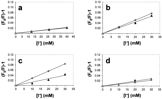
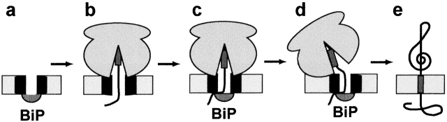
In mammalian cells, most membrane proteins are inserted cotranslationally into the ER membrane at sites termed translocons. Although each translocon forms an aqueous pore, the permeability barrier of the membrane is maintained during integration, even when the otherwise tight ribosome-translocon seal is opened to allow the cytoplasmic domain of a nascent protein to enter the cytosol. To identify the mechanism by which membrane integrity is preserved, nascent chain exposure to each side of the membrane was determined at different stages of integration by collisional quenching of a fluorescent probe in the nascent chain. Comparing integration intermediates prepared with intact, empty, or BiP-loaded microsomes revealed that the lumenal end of the translocon pore is closed by BiP in an ATP-dependent process before the opening of the cytoplasmic ribosome-translocon seal during integration. This BiP function is distinct from its previously identified role in closing ribosome-free, empty translocons because of the presence of the ribosome at the translocon and the nascent membrane protein that extends through the translocon pore and into the lumen during integration. Therefore, BiP is a key component in a sophisticated mechanism that selectively closes the lumenal end of some, but not all, translocons occupied by a nascent chain. By using collisional quenchers of different sizes, the large internal diameter of the ribosome-bound aqueous translocon pore was found to contract when BiP was required to seal the pore during integration. Therefore, closure of the pore involves substantial conformational changes in the translocon that are coupled to a complex sequence of structural rearrangements on both sides of the ER membrane involving the ribosome and BiP.


Figures

Figure 1.
Figure 1.
Three possible mechanisms for sealing the lumenal side of the aqueous translocon pore during protein integration. (a) A conformational change in the translocon prevents ion flow through the pore from the lumenal end. (b) Disassembly of the translocon machinery maintains the permeability barrier of the membrane by eliminating the pore. (c) A soluble lumenal protein mediates, either directly or indirectly, the closure of the lumenal side of the aqueous translocon pore.
Figure 2.
Figure 2.
111p protein and integration intermediates. (a) The chimeric 111p single-spanning membrane protein was constructed using the TM sequence from vesicular stomatitus G protein (residues 65–84, dark gray) and lysine-free stretches of preprolactin and Bcl-2 (Liao et al., 1997). 111p contains only a single lysine codon at position 75 in the middle of the TM sequence (white circle). Note that the TM segment will still be nonpolar and uncharged when ɛNBD-Lys is incorporated at this location. The signal sequence (SS) is indicated in gray (residues 1–22), and the triangle indicates the approximate position of the truncations used in this study (residues 86, 91, and 93). (b) Accessibility of the fluorescent probe from either the cytoplasmic or lumenal sides of intact ER microsomes is indicated for each intermediate used in this study (Liao et al., 1997). The ribosome is shown bound to the translocon (black) at the ER membrane (light gray) in each case. The TM sequence is represented by a dark gray rectangle and the fluorescent probe by a white circle.
Figure 3.
Figure 3.
Iodide ion quenching of NBD-labeled 111p-91 integration intermediates prepared with various ER microsomes. Samples containing NBD-111p-91 integration intermediates prepared with (a) KRMs, (b) XRMs, (c) XRMs + αSec61α, or (d) RRMs (XRMs reconstituted with rBiP) were divided into four equal aliquots. Each sample then received the same total concentration of KCl and KI, but different amounts of KI as shown. Fo is the net fluorescence intensity in the absence of quencher, whereas F is the net fluorescence intensity at a given iodide ion concentration. Measurements were made in the absence (▴) or the presence (•) of pore-forming PFO toxin to introduce iodide ions into the lumen of the microsomes. The linear least-squares best-fit lines for data averaged from several independent experiments are shown. The results are also reported in Tables I and II.
Figure 4.
Figure 4.
Mechanism for maintaining the permeability barrier of the ER membrane during cotranslational membrane protein integration. (a) Prior to integration, the ribosome-free translocon is sealed on the lumenal side by BiP. (b) After SRP-dependent targeting of a ribosome-nascent chain complex to the translocon and translation to yield a nascent chain longer than 70 amino acids (Crowley et al., 1994), the ribosome–translocon seal is intact and the lumenal end of the pore is open (e.g., 111p-86). (c) After the TM sequence has been synthesized and is still near the peptidyltransferase center far inside the ribosome (Liao et al., 1997), the lumenal end of the translocon pore is closed by the action of BiP (111p-88, 111p-91). Although BiP is shown here physically plugging the pore, BiP may effect closure indirectly by binding to another protein(s) that physically closes the pore. At this point, the ribosome–translocon seal is still intact. (d) The ribosome–translocon seal is then broken, whereas the BiP-dependent seal at the other end of the pore remains intact (111p-93). Although the ribosome is depicted here as rotating relative to the translocon, the nature and magnitude of this structural change is not yet known. (e) After termination of translation, the TM sequence is integrated into the ER membrane.

References

    1. Beckmann, R., D. Bubeck, R. Grassucci, P. Penczek, A. Verschoor, G. Blobel, and J. Frank. 1997. Alignment of conduits for the nascent polypeptide chain in the ribosome-Sec61 complex. Science. 278:2123–2126. - PubMed
    1. Beckmann, R., C.M.T. Spahn, N. Eswar, J. Helmers, P.A. Penczek, A. Sali, J. Frank, and G. Blobel. 2001. Architecture of the protein-conducting channel associated with the translating 80S ribosome. Cell. 107:361–372. - PubMed
    1. Bell, C.E., and D. Eisenberg. 1996. Crystal structure of diphtheria toxin bound to nicotinamide adenine dinucleotide. Biochemistry. 35:1137–1149. - PubMed
    1. Brodsky, J.L. 1996. Post-translational protein translocation: not all hsc70s are created equal. Trends Biochem. Sci. 21:122–126. - PubMed
    1. Brodsky, J.L., and A.A. McCracken. 1999. ER protein quality control and proteasome-mediated protein degradation. Semin. Cell Dev. Biol. 10:507–513. - PubMed

Publication types

MeSH terms