. 2002 Jan 21;156(2):377-87.
doi: 10.1083/jcb.200108030. Epub 2002 Jan 21.

The integrin cytoplasmic domain-associated protein ICAP-1 binds and regulates Rho family GTPases during cell spreading

Affiliations

The integrin cytoplasmic domain-associated protein ICAP-1 binds and regulates Rho family GTPases during cell spreading

Simona Degani et al. J Cell Biol. .

Abstract

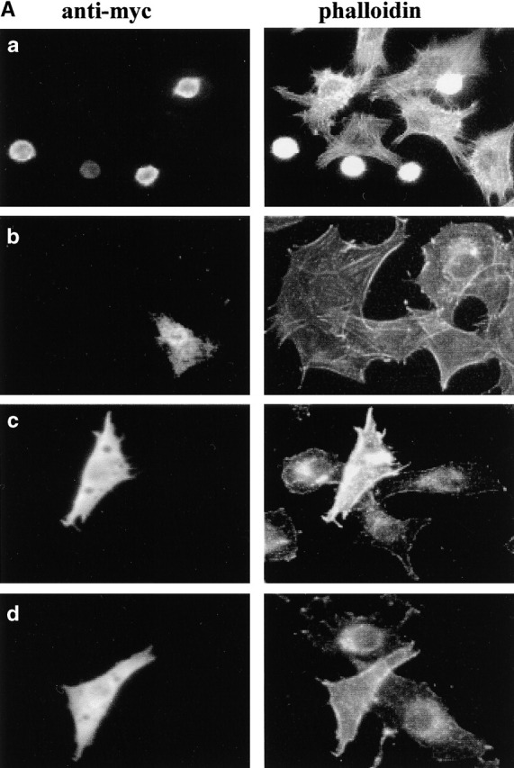
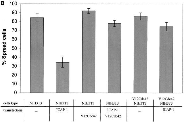
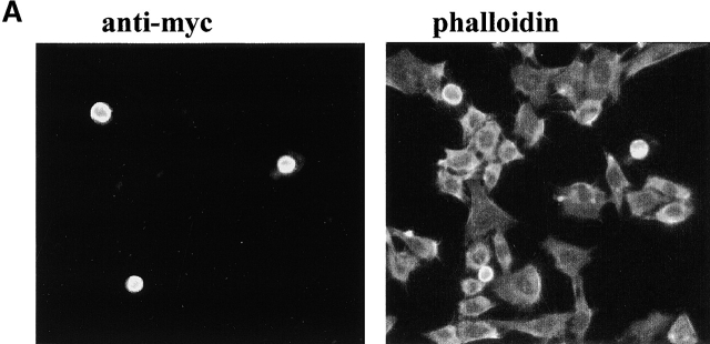
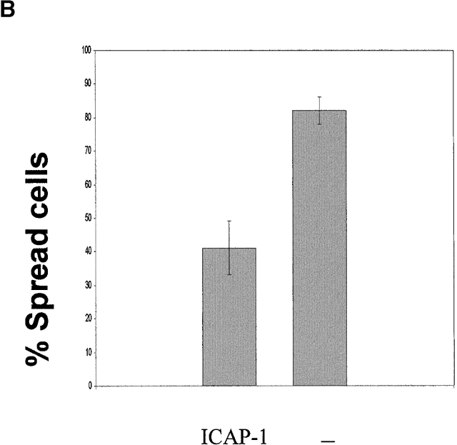
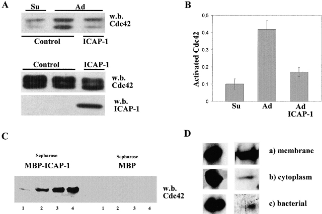
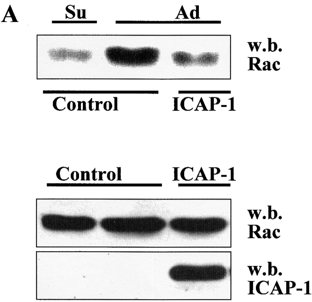
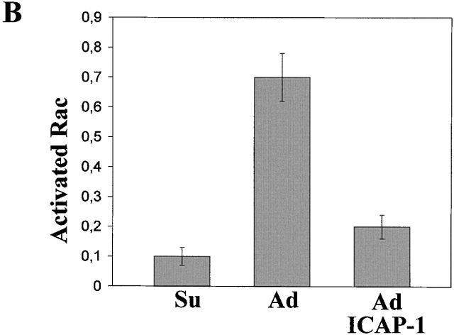
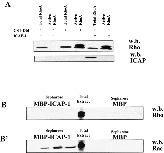
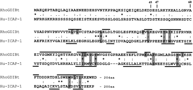
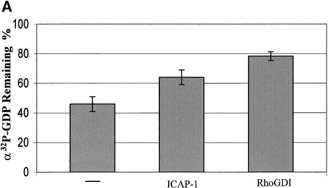
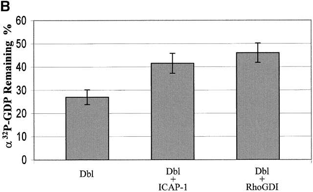
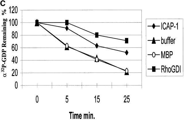
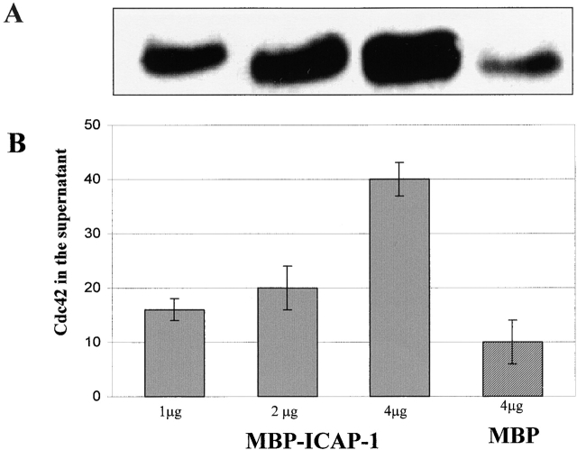
Using two-hybrid screening, we isolated the integrin cytoplasmic domain-associated protein (ICAP-1), an interactor for the COOH terminal region of the beta1A integrin cytoplasmic domain. To investigate the role of ICAP-1 in integrin-mediated adhesive function, we expressed the full-length molecule in NIH3T3 cells. ICAP-1 expression strongly prevents NIH3T3 cell spreading on extracellular matrix. This inhibition is transient and can be counteracted by coexpression of a constitutively activated mutant of Cdc42, suggesting that ICAP-1 acts upstream of this GTPase. In addition, we found that ICAP-1 binds both to Cdc42 and Rac1 in vitro, and its expression markedly inhibits activation of these GTPases during integrin-mediated cell adhesion to fibronectin as detected by PAK binding assay. In the attempt to define the molecular mechanism of this inhibition, we show that ICAP-1 reduces both the intrinsic and the exchange factor-induced dissociation of GDP from Cdc42; moreover, purified ICAP-1 displaces this GTPase from cellular membranes. Together, these data show for the first time that ICAP-1 regulates Rho family GTPases during integrin-mediated cell matrix adhesion, acting as guanine dissociation inhibitor.


Figures

Figure 1.
Figure 1.
ICAP-1 inhibition of cell spreading and rescue by V12Cdc42. Cells were transiently transfected, detached with EDTA, plated on fibronectin-coated coverslips (5 μg/ml) in serum-free medium, fixed, and stained with different antibodies. (A) NIH3T3 cells transfected with myc–ICAP-1 and plated on fibronectin for 1 h (a) and for 24 h (b). NIH3T3 cotransfected with myc–ICAP-1 and myc-V12Cdc42 plated for 1 h on fibronectin (c). V12Cdc42-NIH3T3 transiently transfected with myc–ICAP-1 and plated for 1 h on fibronectin (d). Transfected cells were visualized by anti-myc monoclonal antibody (left), whereas actin organization was detected in the same sample with FITC phalloidin (right). (B) The percentage of spread cells was calculated as described in Materials and methods. Values are means ± SD from three independent experiments in which >20 cells per condition were scored.
Figure 1.
Figure 1.
ICAP-1 inhibition of cell spreading and rescue by V12Cdc42. Cells were transiently transfected, detached with EDTA, plated on fibronectin-coated coverslips (5 μg/ml) in serum-free medium, fixed, and stained with different antibodies. (A) NIH3T3 cells transfected with myc–ICAP-1 and plated on fibronectin for 1 h (a) and for 24 h (b). NIH3T3 cotransfected with myc–ICAP-1 and myc-V12Cdc42 plated for 1 h on fibronectin (c). V12Cdc42-NIH3T3 transiently transfected with myc–ICAP-1 and plated for 1 h on fibronectin (d). Transfected cells were visualized by anti-myc monoclonal antibody (left), whereas actin organization was detected in the same sample with FITC phalloidin (right). (B) The percentage of spread cells was calculated as described in Materials and methods. Values are means ± SD from three independent experiments in which >20 cells per condition were scored.
Figure 2.
Figure 2.
ICAP-1 inhibition of cell spreading on vitronectin. (A) NIH3T3 cells were transiently transfected with myc–ICAP-1, detached with EDTA, and allowed to attach on vitronectin-coated coverslips (10 μg/ml) in serum-free medium for 1 h. Transfected cells were visualized by anti-myc monoclonal antibody, whereas actin organization was detected in the same sample with FITC phalloidin. (B) The percentage of spread cells was calculated as described in Materials and methods. Values are means ± SD from three independent experiments in which >20 cells per condition were scored.
Figure 2.
Figure 2.
ICAP-1 inhibition of cell spreading on vitronectin. (A) NIH3T3 cells were transiently transfected with myc–ICAP-1, detached with EDTA, and allowed to attach on vitronectin-coated coverslips (10 μg/ml) in serum-free medium for 1 h. Transfected cells were visualized by anti-myc monoclonal antibody, whereas actin organization was detected in the same sample with FITC phalloidin. (B) The percentage of spread cells was calculated as described in Materials and methods. Values are means ± SD from three independent experiments in which >20 cells per condition were scored.
Figure 3.
Figure 3.
ICAP-1 inhibits and binds Cdc42. (A) COS cells transiently transfected with myc-Cdc42 together with control vector or myc–ICAP-1 were suspended by trypsin treatment, kept in suspension 2 h 30 min in serum-free DME (Su), and plated on fibronectin (5 μg/ml) for 1 h (Ad). Cdc42 activity assay was performed as described in Materials and methods using a GST-PAK-CD fusion protein that selectively binds to GTP-Cdc42. The bound GTP-Cdc42 was analyzed by Western blotting with a polyclonal antibody to Cdc42. To probe for Cdc42 and ICAP-1 expression, total cell lysates were blotted with the corresponding antibodies. (B) The amount of GTP-bound and total Cdc42 (A, bottom band of the doublets) was quantified by densitometric analysis, and the activation level of Cdc42 was expressed as a ratio between the values of GTP-bound and total Cdc42. Comparable results were obtained in three independent experiments. (C) Total protein extract of COS cells transfected with GST-Cdc42 was loaded on Sepharose coupled with MBP–ICAP-1 fusion protein or MBP as control. The proteins eluted with glycine-HCl, pH 3.0, buffer (fractions numbers 1-2-3-4) were analyzed by Western blotting with polyclonal antibody against Cdc42. Note that in A, Cdc42 is detected as a doublet of bands; comparison of the electrophoretic mobility indicates that the bottom band of the doublet in A comigrates with the band in C. The top band of the doublet is likely to represent an incompletely processed form of the GTPase. (D) Western blotting with an antibody against Cdc42 of the pooled fractions eluted from MBP–ICAP-1-Sepharose columns (right). The columns were loaded with equal amounts of GST-Cdc42 fusion protein obtained from either the membrane fraction of pCEFL-GST-Cdc42–transfected COS cells (a), the cytoplasmic fraction of the same cells (b), or bacterial lysate of E. coli producing the GST-Cdc42 fusion protein (c). The amount of fusion proteins loaded on the column are shown on the left as control for equal loading.
Figure 4.
Figure 4.
ICAP-1 inhibits and binds Rac1. (A) COS cells transiently transfected with control vector or myc–ICAP-1 were suspended by trypsin treatment, kept in suspension 2 h 30 min in serum-free DME (Su), and plated on fibronectin (5 μg/ml) for 1 h (Ad). Rac1 activation level was evaluated as described in Materials and methods using a GST-PAK-CD fusion protein that selectively binds to GTP-Rac1. The bound GTP-Rac1 was analyzed by Western blotting with a polyclonal antibody to Rac1. To probe for Rac1 and ICAP-1 expression, total cell lysates were blotted with the corresponding antibodies. (B) The amount of GTP-bound and total Rac1 (A) was quantified by densitometric analysis; the activation level of Rac1 is expressed as a ratio between the values of GTP-bound and total Rac1. Comparable results were obtained in three independent experiments. (C) Total lysate of COS cells transfected with myc-Rac1 was loaded on Sepharose coupled with MBP–ICAP-1 fusion protein or MBP as control. The proteins eluted with glycine-HCl, pH 3, buffer (fractions numbers 1-2-3-4) were analyzed by Western blotting with a monoclonal antibody against Rac1.
Figure 4.
Figure 4.
ICAP-1 inhibits and binds Rac1. (A) COS cells transiently transfected with control vector or myc–ICAP-1 were suspended by trypsin treatment, kept in suspension 2 h 30 min in serum-free DME (Su), and plated on fibronectin (5 μg/ml) for 1 h (Ad). Rac1 activation level was evaluated as described in Materials and methods using a GST-PAK-CD fusion protein that selectively binds to GTP-Rac1. The bound GTP-Rac1 was analyzed by Western blotting with a polyclonal antibody to Rac1. To probe for Rac1 and ICAP-1 expression, total cell lysates were blotted with the corresponding antibodies. (B) The amount of GTP-bound and total Rac1 (A) was quantified by densitometric analysis; the activation level of Rac1 is expressed as a ratio between the values of GTP-bound and total Rac1. Comparable results were obtained in three independent experiments. (C) Total lysate of COS cells transfected with myc-Rac1 was loaded on Sepharose coupled with MBP–ICAP-1 fusion protein or MBP as control. The proteins eluted with glycine-HCl, pH 3, buffer (fractions numbers 1-2-3-4) were analyzed by Western blotting with a monoclonal antibody against Rac1.
Figure 5.
Figure 5.
ICAP-1 does neither inhibit nor bind RhoA. (A) RhoA activity assay was performed as described in Materials and methods using COS cells either untransfected or transfected as indicated. The same blot was reprobed for ICAP-1 expression with a polyclonal antibody against ICAP-1. (B) Total protein extract of COS cells was loaded on Sepharose coupled with MBP–ICAP-1 fusion protein or MBP as control. The eluted proteins were analyzed by Western blotting with a monoclonal antibody against RhoA. The same filter was stripped and tested for the presence of Rac1 using a specific monoclonal antibody (B'). Total extract is shown as control.
Figure 8.
Figure 8.
Sequence alignment of bovine RhoGDI (RhoGDIBt) and ICAP-1 proteins. Sequences of human ICAP-1 (sequence data available from GenBank/EMBL/DDBJ under accession no. NP_004754) and bovine RhoGDI (accession no. CAA36916) were aligned using CLUSTAL W (1.81). Lines above RhoGDIBt sequence indicate secondary structure elements as reported by Hoffman et al. (2000). Lines under ICAP-1A sequence indicate a prediction of its secondary structure elements as obtained using a Biology Work Bench protein analysis tool based on the DSC algorithm (King and Sternberg, 1990). Shaded grey areas indicate residues of the RhoGDIBt that interact with Cdc42 geranylgeranyl moiety. Asterisks indicate single fully conserved residue. Colons indicate conservation of strong groups. Periods indicate conservation of weak groups.
Figure 6.
Figure 6.
Inhibitory effect of ICAP-1 on the dissociation of [α-32P]GDP from Cdc42. GST-Cdc42 was expressed in COS cells, and the protein was purified from the membranous fraction and captured on glutathione-Sepharose. The Cdc42 immobilized on the beads was loaded with guanosine nucleotide by incubation with [α-32P]GTP 25 min at room temperature. The dissociation of the GDP induced by chelating Mg2+ with EDTA (A) or by the GEF activity of Dbl (B) was measured in the presence of either the buffer alone, MBP–ICAP-1(10 μg), or the same amount of GST-Rho GDI fusion proteins after 10 min at room temperature. (C) Time course for [α-32P]GDP dissociation from Cdc42 induced by chelating Mg2+ with EDTA in the presence of either 10 μg of MBP–ICAP-1, GST-Rho GDI, MBP carrier protein, or buffer alone. Results shown are means ± SD of values from three experiments. [α-32P]GDP remaining is expressed as the percentage of [α-32P]GDP bound to Cdc42 after loading.
Figure 6.
Figure 6.
Inhibitory effect of ICAP-1 on the dissociation of [α-32P]GDP from Cdc42. GST-Cdc42 was expressed in COS cells, and the protein was purified from the membranous fraction and captured on glutathione-Sepharose. The Cdc42 immobilized on the beads was loaded with guanosine nucleotide by incubation with [α-32P]GTP 25 min at room temperature. The dissociation of the GDP induced by chelating Mg2+ with EDTA (A) or by the GEF activity of Dbl (B) was measured in the presence of either the buffer alone, MBP–ICAP-1(10 μg), or the same amount of GST-Rho GDI fusion proteins after 10 min at room temperature. (C) Time course for [α-32P]GDP dissociation from Cdc42 induced by chelating Mg2+ with EDTA in the presence of either 10 μg of MBP–ICAP-1, GST-Rho GDI, MBP carrier protein, or buffer alone. Results shown are means ± SD of values from three experiments. [α-32P]GDP remaining is expressed as the percentage of [α-32P]GDP bound to Cdc42 after loading.
Figure 7.
Figure 7.
ICAP-1 stimulated release of Cdc42 from the membranes of COS cells. Membranes from COS cells transfected with myc-Cdc42 were prepared as described in Materials and methods. The membranes were incubated with the indicated amounts of purified MBP–ICAP-1 or MBP alone as a control for 25 min at room temperature. After centrifugation, the supernatant fractions were examined for the presence of Cdc42 using SDS-PAGE and Western blotting with a specific antibody (A). The relative amount of Cdc42 released into supernatant fraction was determined by densitometric scanning analysis (B).

References

    1. Arthur, W.T., L.A. Petch, and K. Burridge. 2000. Integrin engagement suppresses RhoA activity via a c-Src-dependent mechanism. Curr. Biol. 10:719–722. - PubMed
    1. Balzac, F., S.F. Retta, A. Albini, A. Melchiorri, V.E. Koteliansky, M. Geuna, L. Silengo, and G. Tarone. 1994. Expression of beta1B integrin isoform in CHO cells results in a dominant negative effect on cell adhesion and motility. J. Cell Biol. 127:557–565. - PMC - PubMed
    1. Belkin, A.M., N.I. Zhidkova, F. Balzac, F. Altruda, D. Tomatis, A. Maier, G. Tarone, V.E. Koteliansky, and K. Burridge. 1996. Beta1D integrin displaces the beta 1A isoform in striated muscles: localization at junctional structures and signaling potential in nonmuscle cells. J. Cell Biol. 132:211–226. - PMC - PubMed
    1. Belkin, A.M., S.F. Retta, O.Y. Pletjushkina, F. Balzac, L. Silengo, R. Fassler, V.E. Koteliansky, K. Burridge, and G. Tarone. 1997. Muscle beta1D integrin reinforces the cytoskeleton-matrix link: modulation of integrin adhesive function by alternative splicing. J. Cell Biol. 139:1583–1595. - PMC - PubMed
    1. Bouvard, D., and M.R. Block. 1998. Calcium/calmodulin-dependent protein kinase II controls integrin alpha5 beta1-mediated cell adhesion through the integrin cytoplasmic domain associated protein-1alpha. Biochem. Biophys. Res. Commun. 252:46–50. - PubMed

Publication types

MeSH terms