. 2002 Jan 15;62(2):424-32.

Cyclooxygenase-1 is up-regulated in cervical carcinomas: autocrine/paracrine regulation of cyclooxygenase-2, prostaglandin e receptors, and angiogenic factors by cyclooxygenase-1

Affiliations

Cyclooxygenase-1 is up-regulated in cervical carcinomas: autocrine/paracrine regulation of cyclooxygenase-2, prostaglandin e receptors, and angiogenic factors by cyclooxygenase-1

Kurt J Sales et al. Cancer Res. .

Erratum in

  • Cancer Res 2002 Mar 1;62(5):1579. Soeters Roggert P [corrected to Soeters Robbert P]

Abstract

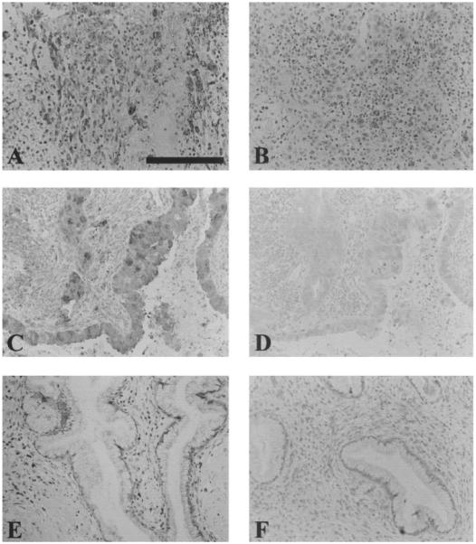
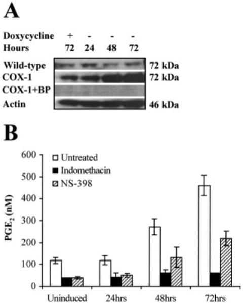
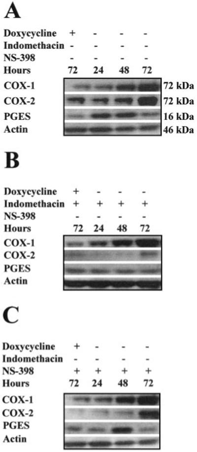
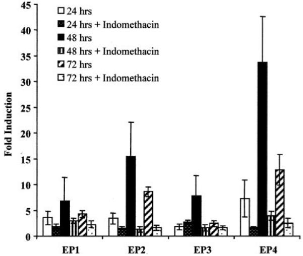
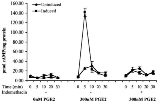
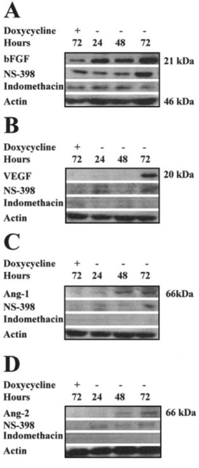
This study was designed to investigate the expression and molecular signaling of cyclooxygenase-1 (COX-1) in cervical carcinomas. Real-time quantitative reverse transcription-polymerase chain reaction and Western blot analysis confirmed enhanced expression of COX-1 RNA, and protein in squamous cell carcinomas and adenocarcinoma of the cervix. COX-1 expression in all carcinoma tissues was associated with enhanced expression of COX-2 RNA and protein. The site of COX-1 expression was localized by immunohistochemistry to the neoplastic epithelial cells in all squamous cell carcinomas and adenocarcinomas studied. Minimal COX-1 immunoreactivity was detected in normal cervix. To explore events associated with COX-1 up-regulation, we developed a doxycycline-regulated expression system in HeLa (cervical carcinoma) cells. Overexpression of COX-1 in HeLa cells resulted in induced expression of cyclooxygenase-2 (COX-2) and prostaglandin E synthase (PGES) concomitant with increased prostaglandin E(2) (PGE(2)) synthesis. Treatment of HeLa cells overexpressing COX-1 with the dual COX enzyme inhibitor indomethacin or selective COX-2 inhibitor NS-398 significantly reduced PGE(2) synthesis. Indomethacin, but not NS-398, treatment abolished the up-regulation of expression of COX-2 and PGES in HeLa cells, suggesting that the observed up-regulation of COX-2 and PGES was mediated by COX-1-enzyme products. To assess whether enhanced PGE(2) synthesis after COX-1 induction would act in an autocrine/paracrine manner, we investigated the effect of COX-1 on the expression of the different isoforms of PGE(2) receptors (EP1-EP4). We found that the cAMP-linked PGE(2) receptors were significantly up-regulated by COX-1 overexpression coincident with enhanced cAMP responsiveness of these cells to exogenous PGE(2) ligand. Finally, overexpression of COX-1 was associated with enhanced expression of the angiogenic factors basic fibroblast growth factor, vascular endothelial growth factor, angiopoietin-1, and angiopoietin-2. This up-regulation of angiogenic factor expression was abolished by indomethacin and partially reduced by NS-398. These data indicate that COX-1 up-regulation modulates the expression of factors that may act in an autocrine/paracrine manner to enhance and sustain tumorigenesis in neoplastic cervical epithelial cells. It is likely that similar mechanisms may act in vivo to modulate tumorigenesis of cervical carcinomas.


Figures

Fig. 1
Fig. 1
A, relative expression of COX-1 and COX-2 RNA in cervical squamous cell carcinoma (C1–C14), adenocarcinoma (C15–C18), and normal cervix (N1–N8) as determined by real-time quantitative RT-PCR. B, Western blot analysis of 50 μg of total protein isolated from human cervical carcinoma tissue. The proteins were loaded onto a 10% SDS-polyacrylamide gel, electrophoresed, and subsequently transferred to a PVDF membrane. The immunoblot was probed with antibody raised against the COOH terminus of human COX-1 or COX-2. A specific band of approximately 72 kDa was detected in all squamous cell carcinomas (panel I; C19C32) and adenocarcinomas (panel II; C33C36). Basal COX-1 expression was detected in seven of eight normal cervices. No COX-2 expression was detected in normal cervical tissue (panel III, N9N16).
Fig. 2
Fig. 2
Localization of COX-1 expression in epithelial cells of squamous cell carcinomas and columnar and glandular epithelium of adenocarcinomas (A and C, respectively). Minimal COX-1 signal was detected in normal cervical tissue (E). Sections that were stained with preadsorbed COX-1 sera are shown in B, D, and F for squamous cell carcinoma, adenocarcinoma, and normal cervix, respectively (negative controls). Scale bar, 100 μm.
Fig. 3
Fig. 3
A, Western blot analysis of 20 μg of total protein isolated from wild-type HeLa Tet-Off and HeLa COX-1 Tet-Off cells grown for 24, 48, and 72 h, respectively, in the absence of DOX. In parallel, control uninduced HeLa COX-1 Tet-Off and wild-type HeLa Tet-Off cells were maintained for 72 h under the same conditions supplemented daily with DOX to a final concentration of 1 μg/ml. The proteins were loaded onto a 4-20% SDS-polyacrylamide gel, electrophoresed, and subsequently transferred to a PVDF membrane. The immunoblot was probed with antibody raised against the COOH terminus of human COX-1. A specific band of approximately 72 kDa was detected. No immunoreactivity was detected by preadsorbing the antibody with the blocking peptide (BP). COX-1 was normalized for protein loading against β-actin on the same blot. B, The functionality of the transfected COX-1 cDNA was assessed by ELISA, by measuring PGE2 secretion into the culture medium after COX-1 induction in the presence or absence of the COX enzyme inhibitor indomethacin, and treatment of HeLa cells with 5 μg/ml arachidonic acid.
Fig. 4
Fig. 4
Western blot analysis of 20 μg of total clarified cell lysate isolated from HeLa COX-1 Tet-Off cells grown for 72 h in the presence of DOX (uninduced) or 24, 48, and 72 h, respectively, in the absence of DOX to induce COX-1 expression. A, expression of COX-2 and PGES was induced coincident with COX-1 overexpression in HeLa cells. B, cotreatment of HeLa cells with indomethacin abolished the COX-1-mediated up-regulation of COX-2 and PGES. C, partial inhibition of the COX-1-mediated up-regulation of COX-2 and PGES expression was observed after cotreatment with the selective COX-2 inhibitor NS-398. Proteins were normalized for loading against β-actin on the same blot.
Fig. 5
Fig. 5
Fold induction of expression of PGE2 receptors (EP1-EP4) in HeLa COX-1 Tet-Off cells as determined by real-time quantitative RT-PCR. COX-1 expression was induced for 24, 48, and 72 h in the presence or absence of the COX enzyme inhibitor indomethacin. Fold induction was determined by dividing the relative expression in induced cells by the relative expression in uninduced cells.
Fig. 6
Fig. 6
cAMP levels in HeLa COX-1 Tet-Off after treatment with 0 or 300 nm PGE2. Cells were either maintained with 1 μg/ml DOX (uninduced) or induced by incubation in culture medium without DOX for 48 h in the presence or absence of the COX enzyme inhibitor indomethacin.
Fig. 7
Fig. 7
Western blot analysis of 20 μg of total clarified cell lysate isolated from HeLa COX-1 Tet-Off cells grown for 72 h in the presence of DOX or for 24, 48, and 72 h in the absence of DOX to induce COX-1 expression. A, immunoblot of bFGF expression after DOX withdrawal from the culture medium. bFGF expression was induced coincident with COX-1 overexpression. Up-regulated bFGF expression was abolished by indomethacin and partially inhibited by NS-398. B, immunoblot of VEGF expression after DOX withdrawal from the culture medium. VEGF was induced after 72 h of COX-1 overexpression. Up-regulated VEGF expression was abolished by indomethacin and partially inhibited by NS-398. C, immunoblot of Ang-1 expression after DOX withdrawal from the culture medium. Ang-1 was induced coincident with COX-1 overexpression after 48 h. Up-regulated Ang-1 expression was abolished by indomethacin and partially inhibited by NS-398. D, immunoblot of Ang-2 expression after DOX withdrawal from the culture medium. Ang-1 was induced after 48 h of COX-1 overexpression. Up-regulated Ang-2 expression was abolished by indomethacin and partially inhibited by NS-398. Proteins were normalized for loading against β-actin.

References

    1. Sitas F, Madhoo J, Wessie J. Incidence of Histologically Diagnosed Cancer in South Africa, 1993-1995. National Cancer Registry of South Africa, South African Institute for Medical Research; Johannesburg: 1998.
    1. Scully RE, Bonfiglio TA, Kurman RJ, Silverberg SG, Wilkinson EJ. Histological Typing of Female Genital Tract Tumors. Springer-Verlag; Berlin: 1994.
    1. DuBois RN, Radhika A, Reddy BS, Entingh AJ. Increased cyclooxygenase-2 levels in carcinogen-induced rat colonic tumors. Gastroenterology. 1996;110:1259–1262. - PubMed
    1. Eberhart CE, Coffey RJ, Radhika A, Giardiello FM, Ferrenbach S, DuBois RN. Up-regulation of cyclooxygenase 2 gene expression in human colorectal adenomas and adenocarcinomas. Gastroenterology. 1994;107:1183–1188. - PubMed
    1. Narko K, Ristimaki A, MacPhee M, Smith E, Haudenschild CC, Hla T. Tumorigenic transformation of immortalized ECV endothelial cells by cyclooxygenase-1 overexpression. J. Biol. Chem. 1997;272:21455–21460. - PubMed

MeSH terms