. 2002 Feb;22(4):979-91.
doi: 10.1128/MCB.22.4.979-991.2002.

Role for the adaptor protein Grb10 in the activation of Akt

Affiliations

Role for the adaptor protein Grb10 in the activation of Akt

Thomas Jahn et al. Mol Cell Biol. 2002 Feb.

Abstract

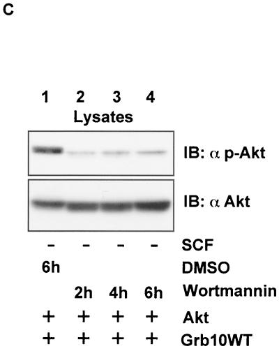
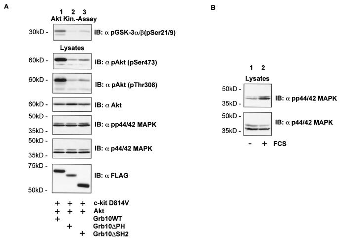
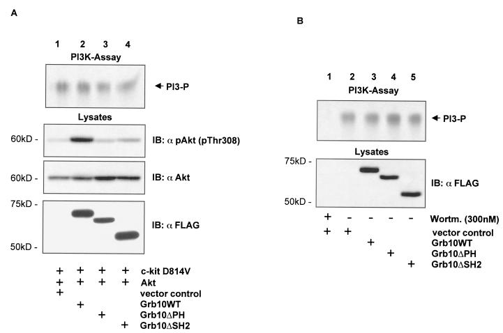
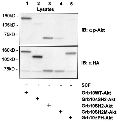
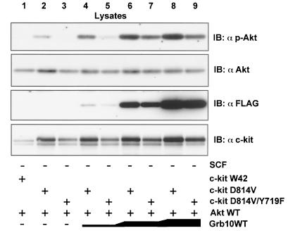
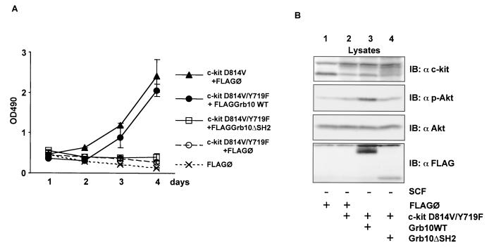
Grb10 is a member of the Grb7 family of adapter proteins lacking intrinsic enzymatic function and encodes functional domains including a pleckstrin homology (PH) domain and an SH2 domain. The role of different Grb10 splice variants in signal transduction of growth factors like insulin or insulin-like growth factor has been described as inhibitory or stimulatory depending on the presence of a functional PH and/or SH2 domain. Performing a yeast two-hybrid screen with the c-kit cytoplasmic tail fused to LexA as a bait and a mouse embryo cDNA library as prey, we found that the Grb10 SH2 domain interacted with the c-kit receptor tyrosine kinase. In the course of SCF-mediated activation of c-kit, Grb10 is recruited to the c-kit receptor in an SH2 domain- and phosphotyrosine-dependent but PH domain-independent manner. We found that Akt and Grb10 form a constitutive complex, suggesting a role for Grb10 in the translocation of Akt to the cell membrane. Indeed, coexpression studies revealed that Grb10 and c-kit activate Akt in a synergistic manner. This dose-dependent effect of Grb10 is wortmannin sensitive and was also seen at a lower level in cells in which c-kit was not expressed. Expression of a Grb10 mutant lacking the SH2 domain as well as a mutant lacking the PH domain did not influence Akt activity. Grb10-induced Akt activation was observed without increased phosphatidylinositol 3-kinase (PI3-kinase) activity, suggesting that Grb10 is a positive regulator of Akt downstream of PI3-kinase. Significantly, deficient activation of Akt by a constitutively activated c-kit mutant lacking the binding site for PI3-kinase (c-kitD814V/Y719F) could be fully compensated by overexpression of Grb10. In Ba/F3 cells, the incapacity of c-kitD814V/Y719F to induce interleukin-3 (IL-3)-independent growth could be rescued by overexpression of Grb10. In contrast, expression of the SH2 deletion mutant of Grb10 together with c-kitD814V/Y719F did not render Ba/F3 cells independent of IL-3. In summary, we provide evidence that Grb10 is part of the c-kit signaling pathway and that the expression level of Grb10 critically influences Akt activity. We propose a model in which Grb10 acts as a coactivator for Akt by virtue of its ability to form a complex with Akt and its SH2 domain-dependent translocation to the cell membrane.


Figures

FIG. 1.
FIG. 1.
Binding of GST-Grb10 SH2 to c-kit. (A) Bound fractions of GST-Grb10SH2 incubated with lysates from Mo7e cells without (lane 1) and with (lane 2) SCF stimulation and GST alone incubated with a lysate from SCF-stimulated Mo7e cells (lane 3) were separated by SDS-PAGE and immunoblotted (IB) with an anti c-kit antibody. (B) Bound fractions of GST-Grb10SH2 (upper panel) and GST alone (lower panel) incubated with lysates from HMC-1 cells without (lane 1) and with (lane 2) SCF stimulation were separated by SDS-PAGE and immunoblotted with an anti c-kit antibody.
FIG. 2.
FIG. 2.
The migration pattern of Grb10 is SCF and wortmannin sensitive. (A) (Top panel) Lysates from Mo7e cells treated with SCF for the times indicated were separated by SDS-PAGE and immunoblotted (IB) with an anti-Grb10 antibody. (Middle panel) Lysates from nonstimulated and SCF-stimulated Mo7e cells treated with wortmannin as indicated were separated by SDS-PAGE and immunoblotted with an anti-Grb10 antibody. (Bottom panel) An anti-Akt immunoprecipitate (IP) from nonstimulated and SCF-stimulated Mo7e cells was separated by SDS-PAGE and immunoblotted with an anti-Akt antibody. (B) An anti-Grb10 immunoprecipitate (upper panel) and an anti-c-kit immunoprecipitate (lower panel) from nonstimulated HMC-1 cells treated with wortmannin as indicated were separated by SDS-PAGE and immunoblotted with an anti-Grb10 and an anti-PY antibody, respectively.
FIG. 3.
FIG. 3.
SCF-induced complex formation between c-kit and Grb10. (A) An anti-Grb10 immunoprecipitation (IP) (top two panels) and a control immunoprecipitation (bottom panel) using equivalent amounts of nonspecific rabbit anti-mouse IgG were simultaneously performed using lysates from nonstimulated and SCF-stimulated Mo7e cells, and the products were separated by SDS-PAGE, and immunoblotted (IB) with the antibodies indicated. (B) COS1 cells (60-mm plates) transiently expressing SS-EYFP-kit (4 μg) and various FLAG epitope-tagged Grb10 constructs (4 μg) as indicated were treated with SCF or left untreated. An anti-FLAG immunoprecipitation and a control immunoprecipitation using equivalent amounts of nonspecific rabbit anti-mouse IgG (rαm) were simultaneously performed. Cell lysates were separated by SDS-PAGE and immunoblotted with the antibodies indicated (top four panels). Lysates from the same cells were separated by SDS-PAGE and immunoblotted with anti-PY and anti-cKit antibodies (bottom two panels). (C) An anti-c-kit immunoprecipitation was performed using lysates from nonstimulated and SCF-stimulated COS1 cells transiently expressing SS-EYFP-kit (3 μg) and FLAG epitope-tagged Grb10WT (3 μg), separated by SDS-PAGE, and immunoblotted with antibodies as indicated (top three panels). Lysates from the same cells were separated by SDS-PAGE and immunoblotted with the antibody indicated (bottom panel).
FIG. 4.
FIG. 4.
Complex formation of Grb10 and Akt. (A) Products of an anti-Grb10 immunoprecipitation (IP) using lysates from Mo7e cells (left panel) and from K562 cells (right panel) were separated by SDS-PAGE and immunoblotted (IB) with the antibodies indicated (top two panels). A control immunoprecipitation (bottom panels) using nonspecific rαm IgG was simultaneously performed, and the product was immunoblotted with α-Akt. (B) Lysates from nonstimulated COS1 cells transiently expressing HA epitope-tagged WT Akt (4 μg) and various FLAG epitope-tagged Grb10 constructs (4 μg) were subjected to an anti-FLAG immunoprecipitation (top two panels) and an anti-HA immunoprecipitation (bottom two panels). A control immunoprecipitation with nonspecific rabbit rαm IgG was performed (third panel from top). Immunoprecipitates were separated by SDS-PAGE and immunoblotted with the antibodies indicated. Lysates of the same cells were separated by SDS-PAGE and immunoblotted with an anti-Akt antibody to confirm equal expression levels of Akt (fourth panel from top). (C) SCF-induced association between c-kit and Grb10 and constitutive association between Grb10 and Akt are independent of Akt activity. An anti-FLAG immunoprecipitation (lanes 5 to 8) and a control immunoprecipitation (lanes 1 to 4) using nonspecific rαm IgG were performed using lysates from COS1 cells transfected with SS-EYFP-kitWT (4 μg), WT Akt (2 μg), and FLAG-tagged Grb10WT (4 μg). The cells were treated with SCF and PI3-K inhibitors prior to lysis as indicated. Immunoprecipitates were separated by SDS-PAGE and immunoblotted with the antibodies as indicated (top three panels). Lysates of the same cells were separated by SDS-PAGE and immunoblotted with a phosphospecific anti-Akt antibody to confirm the activation status of Akt and with an anti-Akt antibody to confirm equal expression levels of Akt. (D) GST-Akt binds to Grb10 and Grb10ΔPH. After preaclearing the lysates from COS1 cells expressing FLAG-tagged Grb10WT (lane 1) and Grb10ΔPH (lane 2) with glutathione beads, 2 μg of GST-Akt or only GST bound to glutathione beads was incubated at 4°C in equal volumes of lysates for 1 h. The beads were washed, and bound fractions were subjected to SDS-PAGE and transferred to PVDF membranes. Bound Grb10 protein was visualized by Western blotting using an anti-FLAG antibody. Lane 3 contains equal amounts of the GST-Akt protein to control cross-reactivity of the anti-FLAG antibody with GST-Akt fragments. This figure shows the results of two separate experiments.
FIG. 5.
FIG. 5.
(A) Coexpression of constitutively activated c-kit and Grb10 reveals SH2 and PH domain-dependent synergism in Akt activation in a concentration-dependent manner. (B) Grb10 alone enhances Akt activity in a concentration-dependent manner. (C) The Grb10 effect on Akt activity is wortmannin dependent. Lysates from COS1 cells (35-mm plates) transiently expressing SS-EYFP-kit D814V (1.5 μg [A]), HA-Akt (1.5 μg [A] and 3 μg [B and C]) and FLAG-Grb10 constructs (increasing amounts of 1, 2, and 3 μg [A and B] and 3 μg [C]) as indicated and treated with inhibitors (C) as indicated were separated by SDS-PAGE and immunoblotted (IB) with the antibodies indicated. DMSO, dimethyl sulfoxide.
FIG. 5.
FIG. 5.
(A) Coexpression of constitutively activated c-kit and Grb10 reveals SH2 and PH domain-dependent synergism in Akt activation in a concentration-dependent manner. (B) Grb10 alone enhances Akt activity in a concentration-dependent manner. (C) The Grb10 effect on Akt activity is wortmannin dependent. Lysates from COS1 cells (35-mm plates) transiently expressing SS-EYFP-kit D814V (1.5 μg [A]), HA-Akt (1.5 μg [A] and 3 μg [B and C]) and FLAG-Grb10 constructs (increasing amounts of 1, 2, and 3 μg [A and B] and 3 μg [C]) as indicated and treated with inhibitors (C) as indicated were separated by SDS-PAGE and immunoblotted (IB) with the antibodies indicated. DMSO, dimethyl sulfoxide.
FIG. 6.
FIG. 6.
(A) Grb10 induces Akt kinase activity. An Akt kinase assay was performed using COS1 cells expressing c-kit D814V (2 μg), HA-Akt (2 μg) or FLAG-Grb10 constructs (4 μg) as indicated. Aliquots of the kinase reaction mixture were separated by SDS-PAGE, and blots were analyzed with an pGSK3α/β antibody (top panel). This assay was performed twice, and similar results were obtained. Lysates from the same cells were separated by SDS-PAGE, and Western blot analyses (IB) were performed using the antibodies indicated. (B) To demonstrate the functionality of the phosphospecific MAPK antibody, COS1 cells were either serum starved for 4 h or grown normally in media containing 10% FCS. Lysates from the cells were separated by SDS-PAGE and immunoblotted with the phosphospecific MAPK antibody (upper panel) or the anti-MAPK antibody (lower level).
FIG. 7.
FIG. 7.
(A) Grb10 induces Akt activity without affecting PI3-K activity. A PI3-K assay was performed using COS1 cells expressing c-kit D814V (3 μg), HA-Akt (3 μg), and FLAG-Grb10 constructs or FLAG vector control (6 μg) as indicated. Equal volumes of lipids from the organic phase were spotted on thin-layer chromatography plates and separated by chromatography. The plates were dried, and radiolabeled lipids were visualized by autoradiography (top panel). Lysates from the same cells were separated by SDS-PAGE, and Western blots analyses (IB) were performed using the antibodies indicated. Three independent experiments of the PI3-K assay showed similar results. (B) PI3-K activity levels (upper panel) were determined from COS1 cells expressing vector control (lanes 1 and 2) either treated for 1 h with wortmannin (300 nM) (lane 1) or untreated (lane 2) and from COS1 cells expressing Grb10 constructs as indicated (lanes 3 to 5). Lysates of the same cells were separated by SDS-PAGE, and Western blot analyses were performed using an anti-FLAG antibody to verify expression of Grb10 constructs (lower panel).
FIG. 8.
FIG. 8.
Fusion constructs of WT Akt with various Grb10 fragments suggest a crucial role of the Grb10 SH2 and PH domains in the activation of Akt. Fusion constructs of HA-tagged WT Akt and various Grb10 constructs were created (see Materials and Methods for details) and transiently expressed in COS1 cells (5 μg). Lysates were separated by SDS-PAGE and immunoblotted (IB) with the antibodies indicated.
FIG. 9.
FIG. 9.
Grb10 rescues impaired Akt activation by c-kitD814V/Y719F. Lysates from COS1 cells transiently expressing various SS-EYFP-kit (2 μg), HA-Akt (3 μg), and FLAG-Grb10 constructs (increasing amounts of 1, 2, and 3 μg) as indicated and treated with SCF or left untreated were separated by SDS-PAGE and immunoblotted (IB) with the antibodies indicated.
FIG. 10.
FIG. 10.
Grb10 expression overrides incapacity of c-kitD814V/Y719F to induce IL-3-independent growth of Ba/F3 cells. (A) Ba/F3 cells stably expressing c-kit and Grb10 constructs as indicated were plated in media without IL-3, in 96-well plates at 3 × 10⁴ cells per well. After the indicated period, the viable cells in each well were assayed by their ability to transform MTS into a purple formazan. The absorbance of the samples was measured in an ELISA reader at 490 nm (OD490). Values for day 5 are not shown, because cells at high density were already overgrown (c-kit D814V+FLAG/O, c-kit D814V/Y719F+FLAGGrb10 WT). (B) Lysates from Ba/F3 cells assayed in panel A were analyzed by Western blotting (IB) for expression of c-kit (top panel, anti-c-kit) and Grb10 constructs (bottom panel, anti-FLAG). Activation of endogenous Akt was analyzed by probing the lysates with a phosphospecific Akt antibody (second panel), and levels of endogenous Akt were determined with an anti-Akt antibody (third panel).

Similar articles

Cited by

References

    1. Ahmed, N. N., T. F. Franke, A. Bellacosa, K. Datta, M. E. Gonzalez-Portal, T. Taguchi, J. R. Testa, and P. N. Tsichlis. 1993. The proteins encoded by c-akt and v-akt differ in post-translational modification, subcellular localization and oncogenic potential. Oncogene 8:1957-1963. - PubMed
    1. Altomare, D. A., K. Guo, J. Q. Cheng, G. Sonoda, K. Walsh, and J. R. Testa. 1995. Cloning, chromosomal localization and expression analysis of the mouse Akt2 oncogene. Oncogene 11:1055-1060. - PubMed
    1. Altomare, D. A., G. E. Lyons, Y. Mitsuuchi, J. Q. Cheng, and J. R. Testa. 1998. Akt2 mRNA is highly expressed in embryonic brown fat and the AKT2 kinase is activated by insulin. Oncogene 16:2407-2411. - PubMed
    1. Andjelkovic, M., D. R. Alessi, R. Meier, A. Fernandez, N. J. Lamb, M. Frech, P. Cron, P. Cohen, J. M. Lucocq, and B. A. Hemmings. 1997. Role of translocation in the activation and function of protein kinase B. J. Biol. Chem. 272:31515-31524. - PubMed
    1. Bai, R. Y., T. Jahn, S. Schrem, G. Munzert, K. M. Weidner, J. Y. Wang, and J. Duyster. 1998. The SH2-containing adapter protein GRB10 interacts with BCR-ABL. Oncogene 17:941-948. - PubMed

Publication types

MeSH terms

LinkOut - more resources