. 2002 Feb;22(4):1126-39.
doi: 10.1128/MCB.22.4.1126-1139.2002.

Unique motif for nucleolar retention and nuclear export regulated by phosphorylation

Affiliations

Unique motif for nucleolar retention and nuclear export regulated by phosphorylation

Frédéric Catez et al. Mol Cell Biol. 2002 Feb.

Abstract

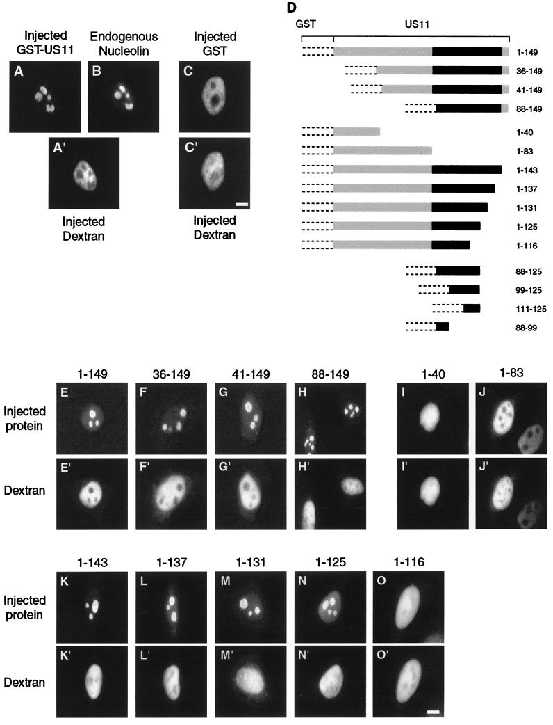
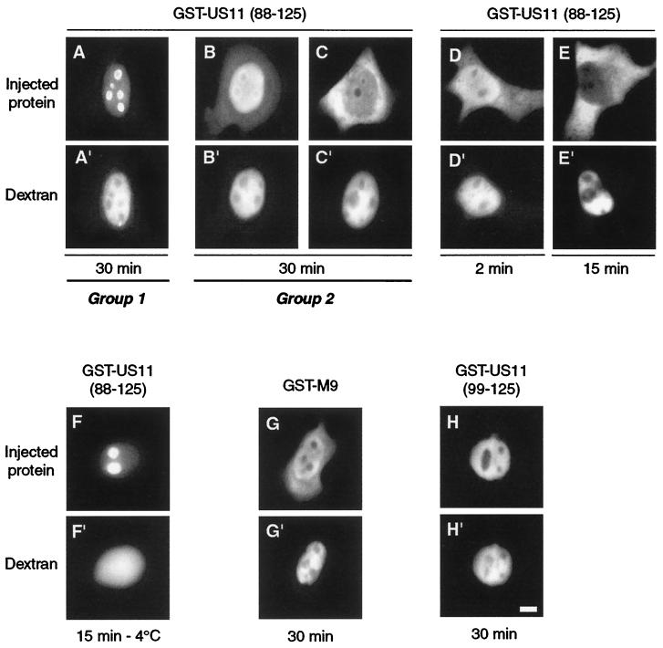
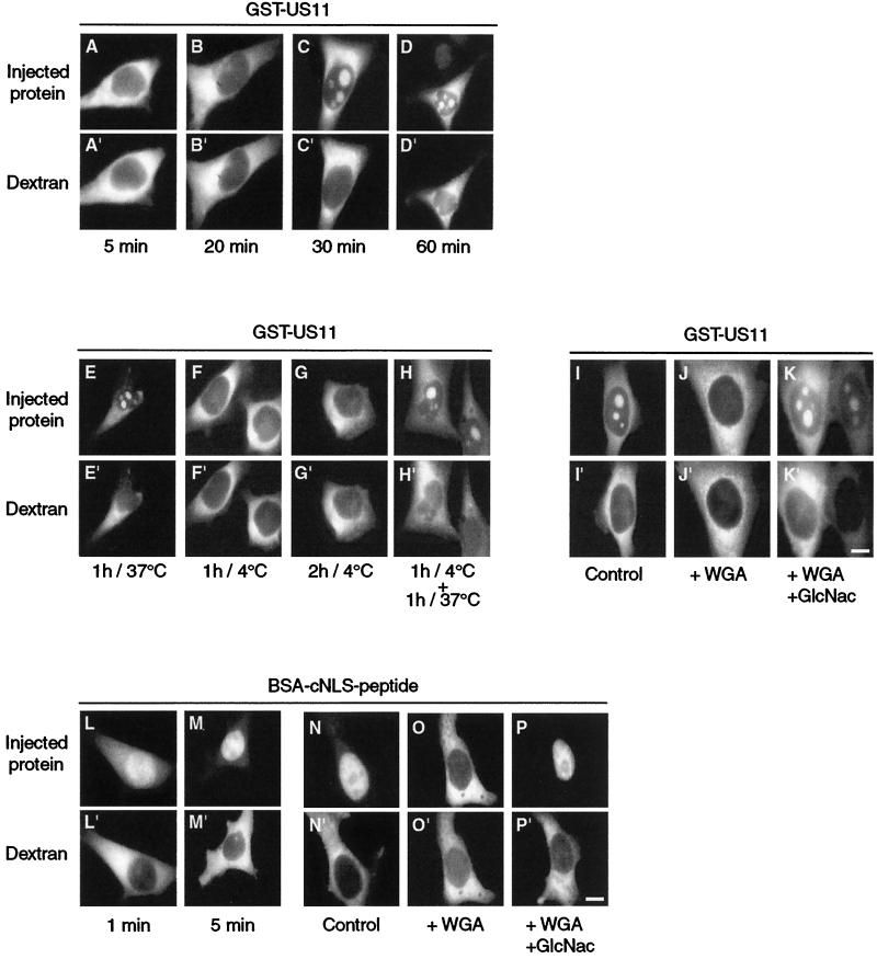
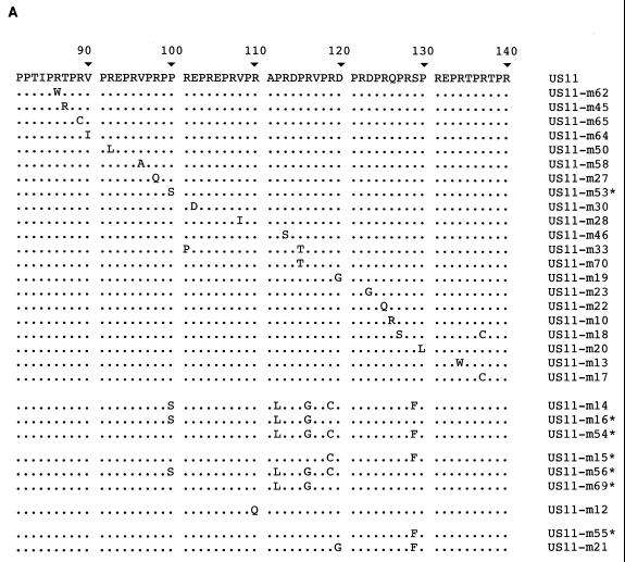
By microinjecting purified glutathione S-transferase linked to all or parts of herpes simplex virus type 1 US11 protein into either the nucleus or the cytoplasm, we have demonstrated that this nucleolar protein exhibits a new type of localization signal controlling both retention in nucleoli and export to the cytoplasm. Saturated mutagenesis combined with computer modeling allowed us to draw the fine-structure map of this domain, revealing a new proline-rich motif harboring both activities, which are temperature dependent and regulated by phosphorylation. Finally, crossing the nuclear pore complex from the cytoplasm to the nucleus is an energy-dependent process for US11 protein, while getting to nucleoli through the nucleoplasm is energy independent.


Figures

FIG. 1.
FIG. 1.
Delineation of US11 NoRS. (A to C) Recombinant hybrid proteins GST-US11 and GST were purified, mixed with high-molecular-weight Texas Red-labeled dextran, and injected into the nuclei of HeLa cells. Injected cells were incubated at 37°C for 30 min and fixed. Injected GST-US11 (A), endogenous nucleolin (B), and injected GST (C) were detected by indirect immunofluorescence using a monoclonal mouse anti-GST antibody (A), a polyclonal rabbit antinucleolin antibody (B), and a rabbit polyclonal anti-GST antibody (C), as well as aminomethylcoumarin acetate-labeled (A) and fluorescein isothiocyanate-labeled (B and C) second antibody. The site of injection was monitored in each injected cell by direct observation of fluorescence of Texas Red-labeled dextran under an inverted microscope (A" and C"). (D) Construction of GST-US11 hybrid proteins. Recombinant hybrid proteins were constructed by cloning parts of the US11 coding sequence from the HSV-1 KOS strain in frame with the GST coding sequence. GST protein is represented by dotted lines, and the US11 coding sequence is represented by a plain box, in which the black part represents the 20 repeats of the XPR sequence. GST-US11 contains the full-length US11 coding sequence. The other hybrid proteins contain the same GST but only parts of the US11 protein. Numbers to the right of each construct indicate the positions of amino acids in the US11 protein. (E to O) Intranuclear localization of GST-US11 hybrid proteins. Recombinant hybrid proteins depicted in D were mixed with Texas Red-labeled dextran and injected into the nuclei of HeLa cells. Hybrid proteins were detected by indirect immunofluorescence 30 min after injection using either an anti-US11 antibody (E to H) or an anti-GST antibody (I to O) and fluorescein isothiocyanate-labeled second antibody. Fluorescence of Texas Red-labeled dextran was directly visualized under an inverted microscope (E" to O"). Bars, 10 μm.
FIG. 2.
FIG. 2.
Identification of a bifunctional NoRS/NES in US11. Three different recombinant hybrid proteins were injected into the nuclei of HeLa cells, GST-US11(88-125) (A to F), GST-US11(99-125) (H), and GST-M9 (G) as a control for nuclear export (see text). Cells were fixed after either 30 min of incubation at 37°C (A to C and G to H), 2 min at 37°C (D), 15 min at 37°C (E), or 15 min at 4°C (F). Hybrid proteins were detected by indirect immunofluorescence using an anti-GST antibody and fluorescein isothiocyanate-labeled second antibody. The two kinds of localization observed (see text for description) are underlined and labeled group 1 and group 2. Fluorescence of Texas Red-labeled dextran was directly visualized under an inverted microscope (A" to H"). Bar, 10 μm.
FIG. 3.
FIG. 3.
Active transport of GST-US11 protein from the cytoplasm to the nucleus. (A to D) Kinetics of nuclear import of GST-US11. Purified GST-US11 protein was mixed with Texas Red-labeled dextran and injected into the cytoplasm of HeLa cells. Cells were fixed after 5, 20, 30, or 60 min of incubation as indicated under each pair of pictures, and GST-US11 was detected by indirect immunofluorescence as described in the legend to Fig. 1. (E to K) Inhibition of nuclear import at 4°C and by WGA. GST-US11 was injected into the cytoplasm, and cells were subsequently incubated at 37 or 4°C for the indicated times (E to G). Injected cells were first incubated at 4°C and then at 37°C for the indicated times (H). GST-US11 was injected either alone (I) or together with WGA (J) or with a mixture of WGA and N-acetylglucosamine (K). (L to P) Nuclear import of BSA-cNLS peptide. As a control for experimental conditions, cNLS conjugated to BSA (see text) was mixed with Texas Red-labeled dextran and injected into the cytoplasm. Kinetics of nuclear import (L and M) and inhibition of nuclear import (N to P) of cNLS-BSA peptide by WGA were analyzed as for that of GST-US11 protein (A to K). BSA-cNLS peptide was detected using fluorescein isothiocyanate-labeled streptavidin. Fluorescence of Texas Red-labeled dextran was directly visualized under an inverted microscope (A" to P"). Bars, 10 μm.
FIG. 4.
FIG. 4.
Modalities of accumulation in nucleoli of GST-US11 protein. (A to D) Kinetics of accumulation in nucleoli after injection into the nucleus. Purified GST-US11 protein was injected into the nucleus and detected by indirect immunofluorescence after 1, 3, 10, or 25 min of incubation at 37°C. (E and G to I) Accumulation in nucleoli of GST-US11 in the absence of energy. HeLa cells were incubated for 30 min at 4°C before injection of GST-US11 into the nuclei of cells, which were maintained at 4°C for another 30 min before fixation (E). HeLa cells were incubated with either rotenone, 2-deoxyglucose, or both for 2 h before measurement of the total cellular ATP. Results are presented as a percentage of the ATP found in control cells (F). GST-US11 was injected into the nuclei of cells treated with rotenone and 2-deoxyglucose. After 30 min at 37°C in the presence of the two compounds, cells were fixed and GST-US11 was detected by indirect immunofluorescence using an anti-US11 antibody. To control the consequence of ATP depletion for the transport of proteins through the NPC, cNLS-BSA peptide was injected into the cytoplasm of cells treated (I) or not (H) with both rotenone and 2-deoxyglucose for 2 h, and its localization was analyzed 15 min after injection using fluorescein isothiocyanate-labeled streptavidin. (J to M) Accumulation in nucleoli of GST-US11 in the absence of transcription. HeLa cells were treated with either 0.05 or 5 μg of actinomycin D (AMD) per ml for 3 h before they were injected with GST-US11 into either the cytoplasm (J and L) or the nucleus (K and M). GST-US11 was detected by indirect immunofluorescence after either 1 h (J and L) or 30 min (K and M) of incubation at 37°C in the presence of the same concentration of actinomycin D. (A" to M") Fluorescence of Texas Red-labeled dextran was directly visualized under an inverted microscope. (N and O) HeLa cells were treated with actinomycin D for 3 h before fixation. (P) Control untreated HeLa cells. In all cases (N to P), endogenous nucleolin was detected by indirect immunofluorescence using an anti-nucleolin antibody. Bars, 10 μm.
FIG. 5.
FIG. 5.
Amino acids involved in NoRS/NES function of US11 protein. (A) Sequences of altered forms of US11 carrying amino acid replacements distributed throughout the domain encompassing the NoRS/NES. The sequence of amino acids 81 to 140 of wild-type US11 protein is reported at the top of the figure. Amino acids are specified by standard single-letter abbreviations. Amino acid sequences from the same region were deduced after sequencing of the 30 different clones of US11 genes and aligned below the wild-type sequence. Amino acids identical to that of the wild-type protein are indicated by dots. Names of the modified forms of US11 are indicated to the right. Nine of the 30 modified US11 proteins exhibited other amino acid replacements or deletions in addition to those shown here, namely, Q for L64 in US11-m45; V for D69 in US11-m65; S for N51 and L for Q57 in US11-m50; E for A55 in US11-m58; S for G41 in US11-m27; L for S73 in US11-m30; M for T144 in US11-m17; and N for I42 and deletion of amino acids 45 to 62 in US11-m33. Seven clones were derived from US11-m14 by a second step of mutagenesis; they are specified by a star after their name. (C to G) Intracellular localization of indicated US11 proteins carrying amino acid substitutions compared to that of wild-type US11 protein (B). Modified US11 proteins were localized by indirect immunofluorescence using an anti-US11 antibody 48 h after transfection. Identical distributions of the different US11 proteins were always obtained whatever the expression level after transfection. Bar, 10 μm.
FIG. 6.
FIG. 6.
Identification of the major phosphorylation site of US11 protein and regulation of NoRS/NES function by Ser 129 phosphorylation. (A) Potential phosphorylation sites of US11. Sequences of the XPR repeats in US11 strain MP, US11 strain KOS, and US11-m55 (strain KOS) are indicated in the single-letter code. Potential phosphorylation sites for MAP kinase are indicated by an arrow, and those for casein kinase II and PKC are indicated by a triangle. A gap in the US11 and US11-m55 sequences of the KOS strains points out 12 additional amino acids present only in US11 of the MP strain (see text). (B and C) Immunoprecipitation of in vivo 32P-labeled US11 protein. US11 was immunoprecipitated with anti-US11 antibody in lysates from control HeLa cells (lanes 1 and 5) and from HeLa cells transfected with either CMV-US11 (MP) (lanes 2 and 6), CMV-US11 (KOS) (lanes 3 and 7), or CMV-US11-m55 (lanes 4 and 8) expression vectors. Immunoprecipitated US11 was subjected to SDS-PAGE and transferred to a nitrocellulose membrane for immunodetection with the same anti-US11 antibody (B). After immunostaining, the membrane shown in B was subjected to autoradiography (C). The position of US11 MP is indicated by a black arrow, and that of US11 KOS is indicated by a grey arrow to the left of panel B. (D to F) Analysis of US11 protein phosphorylation by two-dimensional PAGE. Proteins from lysate of HeLa cells previously transfected with the relevant vectors specified above were extracted and lyophilized for separation by two-dimensional PAGE (see text). Proteins were transferred to a nitrocellulose membrane, and US11 was detected with an anti-US11 antibody as described above (B). The positions of the two phosphorylated derivatives of US11 are indicated by a and b, while that of the unphosphorylated protein is labeled US11. Recombinant hybrid proteins GST-US11(88-131) with Ser (G and I) or Phe (H) in position 129 and GST-US11(88-125) (J) were purified, mixed with Texas Red-labeled dextran, and injected into the nuclei of HeLa cells. Injected cells were incubated at 37°C for 30 min in the presence (I and J) or absence (G and H) of 0.2 μM staurosporine and fixed. Hybrid GST-US11s were detected by indirect immunofluorescence using anti-GST antibody (G to J). Fluorescence of Texas Red-labeled dextran was directly visualized under an inverted microscope (G" to J"). Bar, 10 μm.
FIG. 7.
FIG. 7.
Prediction of the three-dimensional structure of the 88 to 125 domain of US11 protein. An enlarged view of the bipartite NoRS is displayed. The brown color-coded dashed lines show distances compatible with stacking interactions (between 3.6 and 4.9 Å). The NoRS1 and NoRS2 submotifs are represented by the respective 90/95/96 and 111/116/117 residue triads (brown/red/yellow color coding).

Similar articles

Cited by

References

    1. Aitchison, J. D., and M. P. Rout. 2000. The road to ribosomes. Filling potholes in the export pathway. J. Cell Biol. 151:F23-F26. - PMC - PubMed
    1. Andjelkovic, M., S. M. Maira, P. Cron, P. J. Parker, and B. A. Hemmings. 1999. Domain swapping used to investigate the mechanism of protein kinase B regulation by 3-phosphoinositide-dependent protein kinase 1 and Ser473 kinase. Mol. Cell. Biol. 19:5061-5072. - PMC - PubMed
    1. Besse, S., J.-J. Diaz, E. Pichard, K. Kindbeiter, J.-J. Madjar, and F. Puvion-Dutilleul. 1996. In situ hybridization and immunoelectron microscope analyses of the Us11 gene of herpes simplex virus type 1 for transient expression. Chromosoma 104:434-444. - PubMed
    1. Bogerd, H. P., R. E. Benson, R. Truant, A. Herold, M. Phingbodhipakkiya, and B. R. Cullen. 1999. Definition of a consensus transportin-specific nucleocytoplasmic transport signal. J. Biol. Chem. 274:9771-9777. - PubMed
    1. Boulikas, T. 1993. Nuclear localization signals (NLS). Crit. Rev. Eukaryot. Gene Expr. 3:193-227. - PubMed

Publication types

MeSH terms

LinkOut - more resources