. 2002 Feb;22(4):1158-71.
doi: 10.1128/MCB.22.4.1158-1171.2002.

The hematopoiesis-specific GTP-binding protein RhoH is GTPase deficient and modulates activities of other Rho GTPases by an inhibitory function

Affiliations

The hematopoiesis-specific GTP-binding protein RhoH is GTPase deficient and modulates activities of other Rho GTPases by an inhibitory function

Xiaoyu Li et al. Mol Cell Biol. 2002 Feb.

Abstract

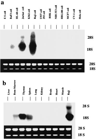
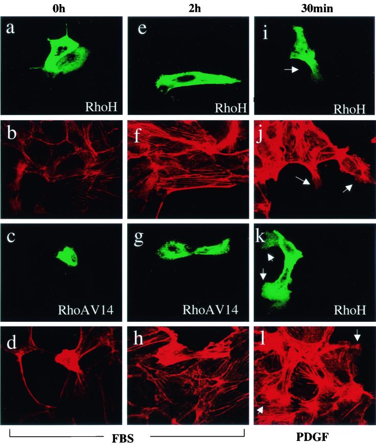
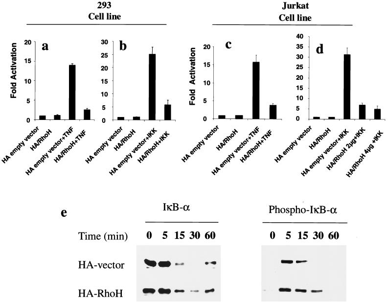
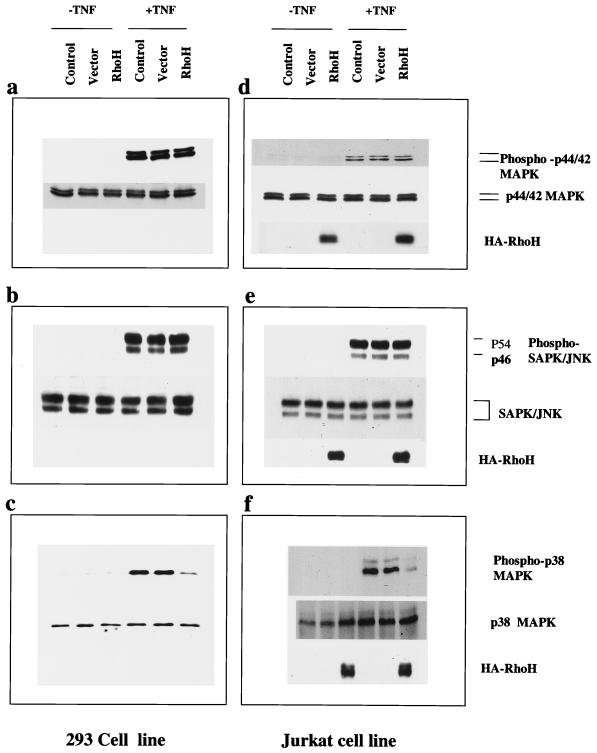
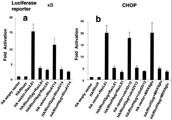
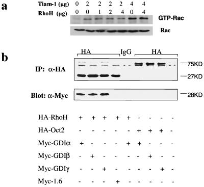
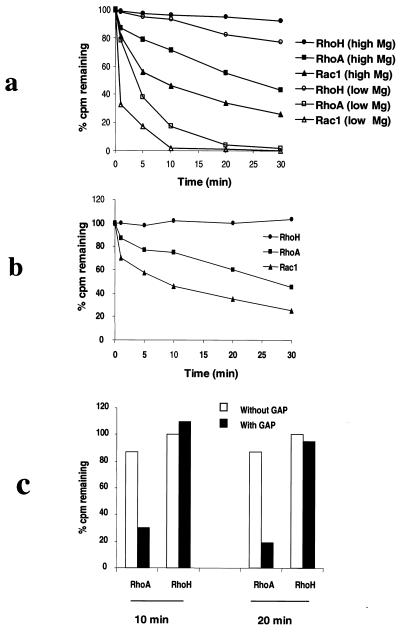
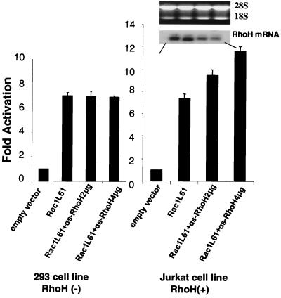
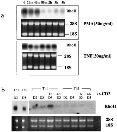
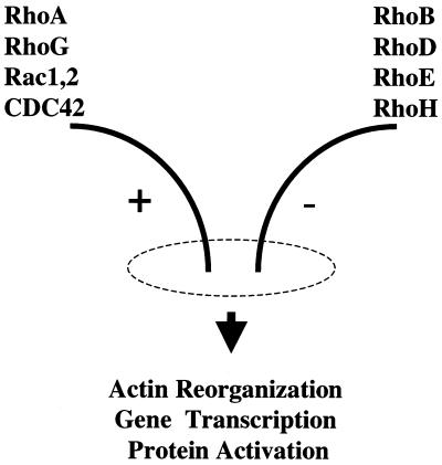
The Rho subfamily of small GTP-binding proteins mediates many fundamental cellular functions. The commonly studied members (Rho, Rac, and CDC42) regulate actin reorganization, affecting diverse cellular responses, including adhesion, cytokinesis, and motility. Another major function of the Rho GTPases is their role in regulating transcriptional factors and nuclear signaling. RhoH is encoded by a hematopoiesis-specific Rho-related gene recently identified in a fusion transcript with bcl6 in lymphoma cell lines. Significantly, translocations and a high frequency of RhoH mutation have been detected in primary lymphoma cells. We show here that RhoH functions differently from other Rho GTPases. RhoH exerts no significant effect on actin reorganization. However, RhoH is a potent inhibitor of the activation of NFkappaB and p38 by other Rho GTPases. This property, together with the differential expression of RhoH in the Th1 subset of T cells, suggests a role for RhoH in the functional differentiation of T cells. RhoH has different amino acids in two highly conserved residues critical for GTPase activity. Consequently, RhoH is GTPase deficient, remaining in a GTP-bound activated state without cycling. Reduction of RhoH levels in T cells augments the response to Rac activation. Furthermore, RhoH is dramatically down regulated after phorbol myristate acetate treatment and in Th1 cells after activation by anti-CD3. Hence, a mechanism for regulation of RhoH function is likely to exist at the transcriptional level. The inhibitory function of RhoH supports a model in which Rho GTPases with opposing functions may compete to modulate the final outcome of a particular GTPase-activated pathway.


Figures

FIG. 1.
FIG. 1.
RhoH is a hematopoiesis-specific gene. (a) Expression in cell lines. RhoH is expressed at high levels in T (Jurkat) and B (Raji) cell lines and at low levels in the mixed lymphoid-erythroid line BaF3. In myeloid (HL60 and U937) and erythroid (K562) cell lines, RhoH is not detectable. All nonhematopoietic cells lines, including undifferentiated embryonal stem (ES) cells, are negative for RhoH. (b) Expression in tissues. mRNA was seen at high levels in the thymus, was less abundant in the spleen, and was lowest in abundance in bone marrow. In all other tissues, RhoH mRNA was nondetectable.
FIG. 2.
FIG. 2.
RhoH has no effect on the cytoskeleton. MDCK cells were transfected with a HA-RhoH or a GST-RhoAV14-expressing vector and, after 24 h of recovery, starved for 48 h. Some cells were fixed and immunostained for RhoH or RhoA (fluorescein isothiocyanate) and actin (rhodamine). Cells were then refed with serum-containing medium (10% FBS) or PDGF (10 ng/ml), fixed at different times after refeeding, and immunostained. (a and b) Starved cells showed minimal stress fibers. RhoH-positive cells did not show any difference in morphology or actin staining pattern. (c and d) RhoAV14-positive cells showed dense stress fibers and cortical actin staining. (e and f) At 2 h after refeeding with 10% FBS medium, increased stress fibers and actin staining were seen in MDCK cells. RhoH-positive cells showed no inhibition of stress fiber formation. (g and h) At 2 h after serum stimulation, RhoV14-positive cells showed even denser actin staining. (i, j, k, and l) At 30 min after restimulation with PDGF, ruffling of cells (arrows) and increased stress fibers were seen. RhoH-positive cells did not show any inhibition of ruffling.
FIG. 3.
FIG. 3.
RhoH inhibits activation of NF-κB. (a and c) An NF-κB-luciferase reporter vector was cotransfected with 2 μg of RhoH into 293 (a) or Jurkat (c) T cells. At 24 h after transfection, cell lysates were evaluated for luciferase activity. RhoH did not induce any increase in luciferase activity above that of an HA-vector control. At 24 h after transfection, cells were treated with TNF. At 6 h after stimulation, maximal luciferase activity (greater than 10-fold induction) was detected in cells transfected with the empty vector, reflecting activation of NF-κB by TNF. In cells transfected with RhoH, NF-κB activation by TNF was almost completely suppressed. (b and d) The pcDNA3IKKβ and NF-κB-reporter vectors were cotransfected with a control vector or RhoH into 293 (b) or Jurkat (d) cells. At 24 h later, cell lysates were evaluated for luciferase activity. IKKβ induced vigorous NF-κB activation (greater than 25-fold induction). In cells transfected with 2 or 4 μg of RhoH, IKK activation of NF-κB was again strongly suppressed with further suppression at a greater RhoH dose. (e) RhoH inhibits IκB degradation but not phosphorylation by IKK. At 24 h after transfection with a control vector or RhoH, 293 cells were treated with TNF. At different times after stimulation, cell lysates were prepared for Western blotting, the level of IκB was measured by anti-IκBα antibody, and the level of phosphorylated IκB was measured by anti-phospho-IκB antibody. Note that in cells transfected with the vector, rapid degradation of IκBα was observed. In cells transfected with RhoH, significantly retarded degradation of IκB was seen and at no time did IκB disappear completely. No difference in phosphorylation of IκB was seen between control vector- and RhoH-transfected cells. Again, there was an obvious retardation of degradation of phosphorylated IκB in RhoH-transfected cells.
FIG. 4.
FIG. 4.
RhoH inhibits p38 but not ERK or JNK activation. 293 or Jurkat cells were transfected with 2 μg of an empty or RhoH vector. At 24 h after recovery, cells were treated with TNF for 30 min and then harvested for Western blot analysis. Activation of ERK, JNK, and p38 was evaluated by anti-phospho-ERK1/ERK2, -JNK, and -p38 antibodies. The expression level of HA-tagged RhoH protein was revealed by anti-HA antibody, which is shown here only for transfected Jurkat cells (d, e, and f). The results show that RhoH did not activate ERK (a and d), JNK (b and e), or p38 (c and f). Cells treated with TNF showed clear activation of JNK, ERK, and p38, as measured by the emergence of the phosphorylated proteins (+TNF). Cells transfected with the control vector showed the same level of activation as nontransfected cells. RhoH did not inhibit activation of ERK (a and d) or JNK (b and e) by TNF. The amount of phospho-p38 was, however, significantly reduced in RhoH-transfected 293 (c) and Jurkat (f) cells.
FIG. 5.
FIG. 5.
RhoH inhibits Rac1, RhoA, and CDC42. Jurkat cells were cotransfected by electroporation with HA-RhoH and constitutively activated RacL61, RhoAV14, or CDC42L61 and assayed for an effect on NF-KB activation (a) or p38 activation (b) by using luciferase reporters as indicated. RacL61 and RhoAV14 induced robust activation of NF-κB. Both activations were effectively suppressed by 2 or 4 μg of RhoH. RacL61, CDC4261L, and MKK6glu induced a vigorous activation of p38 (b) in Jurkat cells that was suppressed by RhoH.
FIG. 6.
FIG. 6.
RhoH does not inhibit exchange factor TIAM-1 and binds to Rho GDP dissociation inhibitors. (a) A 2-μg sample of TIAM-1 was cotransfected with RhoH (1, 2, or 4 μg) by electroporation into Jurkat cells, and 24 h later, the cells were harvested and measured for levels of GTP-Rac with the PBD assay. An increased level of GTP-Rac was seen in TIAM-1-transfected cells. Cotransfection of RhoH did not change the level of GTP-bound Rac. (b) Myc-tagged Rho GDI-α, -β, -γ, and−1.6 (used as a control) were cotransfected into 293 cells with HA-tagged RhoH or HA-tagged Oct2 (used as a control). At 24 h after transfection, cell lysates were prepared from transfected cells and immunoprecipitation (IP) was carried out with anti-HA antibody or immunoglobulin G (used as a control). Immunoprecipitates were separated and transferred to a Western blot. The filter was then probed with anti-HA and anti-myc epitope antibodies. The top of panel b shows the specific immunoprecipitation of HA-RhoH (27 kDa) or HA-Oct2 (75 kDa) protein. Equal levels of protein reflect uniformity of transfection and immunoprecipitation. The lower part of panel b shows the coprecipitation of only Rho GDIs with HA-RhoH. Note the equal levels of Rho GDIs, indicating equal avidity of binding between RhoH and the three different Rho GDIs.
FIG. 7.
FIG. 7.
RhoH has different residues at two key sites that are critical for GTPase activity. Alignment of the amino acid sequences of RhoH, RhoE, RhoA, Rac1, and CDC42 in the GTPase-determining domain. At position 13 of RhoH, a serine is found instead of the glycine at the corresponding position in RhoA, Rac, and CDC 42. At position 62, an asparagine is found in RhoH instead of the glutamine in RhoA, Rac, and CDC42.
FIG. 8.
FIG. 8.
RhoH binds GTP only but has no GTPase activity. (a) Nucleotide dissociation assay using GST-RhoH, -Rac, and -RhoA fusion proteins. Similar to RhoA and Rac1, RhoH binds GTP rapidly upon incubation with radiolabeled nucleotide under nucleotide exchange conditions. Note that RhoH is almost completely resistant to GDP dissociation compared to Rac and RhoA under both high- and low-magnesium conditions. (b) GTP hydrolysis assay: Under conditions that allow RhoA and Rac to autohydrolyze greater than 80% of bound GTP, RhoH did not show any hydrolysis of GTP. (c) RhoH is resistant to Rho GAP p50. Addition of Rho GAP p50 enhanced the hydrolysis activity of RhoA but had no effect on RhoH.
FIG. 9.
FIG. 9.
Antisense blocking of RhoH increases Rac1-induced activity. 293 or Jurkat cells were cotransfected with reporter genes for IFN-γ-Luc (2 μg) and Rac1L61 (2 μg), with or without an αs-RhoH vector (2 or 4 μg). At 24 h after transfection, cell were processed either for luciferase activity measurement or for Northern analysis to assess endogenous levels of RhoH mRNA. (a) In 293 cells, Rac1L61 induced IFN-γ activation and no difference between activity levels was caused by αs-RhoH expression. (b) In Jurkat cells, RacL61 induces an equivalent level of IFN-γ activation. In cells transfected with αs-RhoH, there was an increase in IFN-γ activity proportionate to the reduction in the endogenous RhoH mRNA level. The experiment was repeated two times, and a typical result is shown.
FIG. 10.
FIG. 10.
RhoH is transcriptionally regulated. (a) Jurkat cells were treated with PMA or TNF, and at different time points after stimulation, total RNA was extracted for Northern analysis to assess levels of RhoH mRNA. A 15-μg sample of total RNA was loaded in each lane. PMA was tested at 20, 50, and 100 ng/ml, and TNF was tested at 10, 20, and 50 ng/ml. The results produced by the different treatment dosages are comparable. A typical example is shown. PMA dramatically down regulates RhoH mRNA between 40 and 80 min after treatment. There is little change in RhoH mRNA levels with TNF treatment, except for a small but consistently observed transient decrease at around 40 min. (b) Differential expression in Th1 and Th2 T cells. Naive CD4+ CD62L+ CD44low T cells were separated and treated under Th1 or Th2 differentiating condition as described in Materials and Methods. At days 2 (D2) and 3 (D3) after culture initiation, cells were processed for Northern analysis. (Left) Analysis of 15 μg of total RNA at day 3. The level of RhoH in Th1 cells is about three times as high as that in Th2 cells. (Right) Analysis of 7 μg of total RNAs. Again, the level of RhoH in Th1 cells is higher than that in Th2 cells. At day 3, aliquots of cells were washed and restimulated with anti-CD3. At 1 and 4 h after restimulation, RNA was extracted for Northern analysis. In Th1 cells, down regulation of RhoH was observed by 4 h (the increase at 1 h is likely from a slightly higher level of RNA loading). In Th2 cells, the levels of RhoH remained very low.
FIG. 11.
FIG. 11.
Model of competing Rho GTPases. Two groups of Rho GTPases that have opposite functions are shown as having a positive or negative effect. The model suggests that some cellular end responses may be modulated by competition between these Rho GTPases.

Similar articles

Cited by

References

    1. Adamson, P., C. J. Marshall, A. Hall, and P. A. Tilbrook. 1992. Posttranslational modifications of p21rho proteins. J. Biol. Chem. 267:20033-20038. - PubMed
    1. Adra, C. N., D. Manor, J. L. Ko, S. Zhu, T. Horiuchi, L. Van Aelst, R. A. Cerione, and B. Lim. 1997. RhoGDIgamma: a GDP-dissociation inhibitor for Rho proteins with preferential expression in brain and pancreas. Proc. Natl. Acad. Sci. USA 94:4279-4284. - PMC - PubMed
    1. Bagrodia, S., and R. A. Cerione. 1999. Pak to the future. Trends Cell Biol. 9:350-355. - PubMed
    1. Baldi, L., K. Brown, G. Franzoso, and U. Siebenlist. 1996. Critical role for lysines 21 and 22 in signal-induced, ubiquitin-mediated proteolysis of I kappa B-alpha. J. Biol. Chem. 271:376-379. - PubMed
    1. Baldwin, A. S., Jr. 1996. The NF-kappa B and I kappa B proteins: new discoveries and insights. Annu. Rev. Immunol. 14:649-683. - PubMed

Publication types

MeSH terms

Substances