. 2002 Feb;22(4):1218-32.
doi: 10.1128/MCB.22.4.1218-1232.2002.

The AT-hook protein D1 is essential for Drosophila melanogaster development and is implicated in position-effect variegation

Affiliations

The AT-hook protein D1 is essential for Drosophila melanogaster development and is implicated in position-effect variegation

Nathalie Aulner et al. Mol Cell Biol. 2002 Feb.

Abstract

We have analyzed the expression pattern of the D1 gene and the localization of its product, the AT hook-bearing nonhistone chromosomal protein D1, during Drosophila melanogaster development. D1 mRNAs and protein are maternally contributed, and the protein localizes to discrete foci on the chromosomes of early embryos. These foci correspond to 1.672- and 1.688-g/cm(3) AT-rich satellite repeats found in the centromeric heterochromatin of the X and Y chromosomes and on chromosomes 3 and 4. D1 mRNA levels subsequently decrease throughout later development, followed by the accumulation of the D1 protein in adult gonads, where two distributions of D1 can be correlated to different states of gene activity. We show that the EP473 mutation, a P-element insertion upstream of D1 coding sequences, affects the expression of the D1 gene and results in an embryonic homozygous lethal phenotype correlated with the depletion of D1 protein during embryogenesis. Remarkably, decreased levels of D1 mRNA and protein in heterozygous flies lead to the suppression of position-effect variegation (PEV) of the white gene in the white-mottled (w(m4h)) X-chromosome inversion. Our results identify D1 as a DNA-binding protein of known sequence specificity implicated in PEV. D1 is the primary factor that binds the centromeric 1.688-g/cm(3) satellite repeats which are likely involved in white-mottled variegation. We propose that the AT-hook D1 protein nucleates heterochromatin assembly by recruiting specialized transcriptional repressors and/or proteins involved in chromosome condensation.


Figures
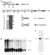
FIG. 1.
FIG. 1.
Developmental expression profile of the D1 gene. (A) Diagrams representing the primary structures of the 11-kDa HMGA1a and the 37-kDa D1 proteins. HMGA proteins contain three AT hooks (I, II, and III) represented by boxes with a hook and a short acidic C-terminal domain (black box). An 11-amino-acid insert generated by alternative splicing of the HMGA mRNA is absent in HMGA1b and present in HMGA1a. The AT hook-like sequences of D1 (5) are numbered from 1 to 10 and are represented by boxes with hooks for full-length or near full-length motifs and by gray boxes for shorter sequences containing the conserved GRP core motif. Acidic C-terminal domains are represented by filled boxes. The corresponding AT hook sequences are shown below the map and can be used to extrapolate a D1 AT hook consensus. The sequence of the human HMGA AT hook consensus is also shown. (B) poly(A)+ mRNA from flies at various developmental stages was hybridized to labeled D1 and RPL17 probes which detect 1.6- and 0.9-kb bands, respectively. poly(A)+ mRNA was purified from 0- to 2-h embryos (lane 1), 0- to 10-h embryos (lane 2), 10- to 20-h embryos (lane 3), first-, second-, and third-instar larvae (lanes 4 to 6, respectively), pupae (lane 7), adult males (lane 8), adult females (lane 9), and dissected ovaries (lane 10). (C) Western blot of protein extracts prepared from Kc cells (lane 1) or 0- to 20-h embryos (lane 2) probed with an antibody raised against the D1 protein.
FIG. 2.
FIG. 2.
The EP473 P-element insertion results in down-regulation of D1. (A) Map of the genomic D1 locus. Sequence coordinates are given relative to the start of the D1 mRNA (+1). Exons are represented by black boxes and 5" and 3" untranslated regions are in gray. Restriction enzyme sites shown are those used to characterize the EP473 P-element insertion and excisions thereof. The P-element insertion diagrammed below the map occurs at position −66. The thick line indicates the extent of the transposon bounded by 5" and 3" inverted repeats (IR; divergent filled arrows). (B) Coomassie blue-stained gel loaded with equivalent amounts of total proteins from wild-type, heterozygous, or homozygous EP473 mutant first-instar larvae and from Kc nuclei (lanes 1 to 4, respectively). A duplicate gel was subjected to Western blot analysis as described above (C). The positions of the D1 bands observed in embryos and tissue-culture cells are indicated to the right. (D) In situ hybridizations to D1 mRNA were performed with wild-type (a to c) and homozygous (d to f) EP473 embryos identified as described in Materials and Methods. (E) Wild-type larvae and homozygous EP473 force-hatched first-instar larvae (see text for details) were processed for DNA staining (red) and D1 immunolabeling (green). Merged signals are shown for wild-type and mutant larval nuclei. Note the smooth appearance of the nuclei in the absence of D1, the smaller space occupied by chromatin and the overall absence of differentially condensed regions of chromatin relative to wild-type nuclei. Bar = 5 μm.
FIG. 3.
FIG. 3.
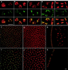
Localization of D1 protein during early embryonic cell cycles and localization in heterochromatin. Embryos at the synctitial preblastoderm stage were processed for confocal microscopy after immunolabeling with the anti-D1 antibody. D1 was revealed using a fluorescein-coupled antibody (green signal) and DNA was stained with propidium iodide (red signal). (a to e) Distribution of D1 as chromosomes progress through the cell cycle (a, interphase; b, early prophase; c, prometaphase; d, early/middle anaphase; e, late anaphase). Bar = 5 μm. Confocal sections taken through embryos during interphase (f to h) or mitosis (i to k) show that D1 localizes exclusively to centromeric heterochromatin at cellularization. Images taken at the apical pole of the embryos (f and i), through the embryos (g and j), and at their periphery (h and k) show that no D1 can be detected in euchromatin. Bar = 5 μm.
FIG. 4.
FIG. 4.
D1 colocalizes with SATI and SATIII repeats in vivo. D1 (red signal) was detected in whole-mount early embryos stained with DAPI (blue) and Lex9F (green). Each row of photographs shows nuclei and chromosomes from embryos at different stages of the cell cycle. The signals shown are identified above each column and illustrate the strict colocalization of D1 with Lex9F, which specifically detects SATI and SATIII repeats. Bars = 5 μm.
FIG. 5.
FIG. 5.
Mapping of D1 binding sites on metaphase chromosomes. Metaphase chromosomes from larval neuroblasts were stained for D1, DNA, and Lex9F as described above. Chromosome sets are shown for both female and male brains. A single interphase nucleus can also be seen. The DAPI, D1, and Lex9F signals shown are as labeled on the photographs. D1 associates with discrete regions of all chromosomes except chromosome 2, consistent with the known localization of SATI and SATIII repeats. The colocalization of D1 with SATIII repeats in the centromeric region of the X chromosome is particularly evident. Bars = 5 μm.
FIG. 6.
FIG. 6.
Dynamic redistribution of D1 during gametogenesis. Adult gonads were stained for DNA and D1 protein and processed for confocal microscopy. (A) Different views of female ovaries: global and more detailed views of the anterior part of an ovariole (a and b, respectively) showing the germarium, ovarian chambers (c and d), and DNA, D1, and merged images of a late-stage oocyte (e, f, and g, respectively). The germarium (ge), follicle cells (fc), nurse cells (nc), the karyosome (k), and oocyte (oo) are indicated. A similar analysis is shown for male testes (B). D1 was revealed by immunofluorescence as described above or by peroxidase staining (d). A diagram adapted from one by Lindsley and Tokuyasu (37) is shown (a) and represents the first half-gyre of a testis with the different spermatocyte cysts that form single-cell layers near the testicular walls. A whole gonad is shown (b) together with a close-up view of the D1 signal in cells from the central part of the testis (top inset). In the germarium (d and e and diagrammed in c), D1 localizes to bright foci in both apical cells (ac) and spermatogonia (sg). Cells situated just below correspond to spermatocytes (sp) in which the protein has redistributed to fill most of the nucleoplasm (e, lower area; f, magnification of panel e).
FIG. 7.
FIG. 7.
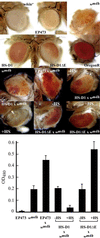
D1 is a modifier of wm4h PEV. (Top two rows) Representative eyes from 5-day-old female parents used in these studies: white, EP473, wm4h, HS-D1, HS-D1ΔE, and a wild-type OregonR stock. The light eye color contributed by the mini-white gene carried by EP473 and the HS-D1 and HS-D1ΔE P elements, ranging from pale yellow to pale orange, did not interfere with analysis of the modification of PEV. (Bottom two rows) Eye color of representative 5-day-old females obtained from the following: wm4h control, EP473 × wm4h, HS-D1 × wm4h without (−HS) or with (+HS) heat shock, and HS-D1ΔE × wm4h without or with heat shock. Flies recovered from the crosses shown in each photograph and in the graph below have the following full genotypes: EP473 × wm4h: wm4h/w; EP473/TM6b. HS-D1 × wm4h: wm4h/w HS-D1. HS-D1ΔE × wm4h: wm4h/w; HS-D1ΔE/+. The genotypes of the parents are indicated in Materials and Methods. Corresponding eye pigment levels (see Materials and Methods) are indicated in the graph below and correspond to mean values ± standard errors from three independent measurements.

References

    1. Alfageme, C. R., G. T. Rudkin, and L. H. Cohen. 1976. Locations of chromosomal proteins in polytene chromosomes. Proc. Natl. Acad. Sci. USA 73:2038-2042. - PMC - PubMed
    1. Alfageme, C. R., G. T. Rudkin, and L. H. Cohen. 1980. Isolation, properties and cellular distribution of D1, a chromosomal protein of Drosophila. Chromosoma 78:1-80. - PubMed
    1. Amirand, C., A. Viari, J. P. Ballini, H. Rezaei, N. Beaujean, D. Jullien, E. Käs, and P. Debey. 1998. Three distinct sub-nuclear populations of HMG-I protein of different properties revealed by colocalization image analysis. J. Cell Sci. 111:3551-3561. - PubMed
    1. Aravind, L., and D. Landsman. 1998. AT-hook motifs identified in a wide variety of DNA-binding proteins. Nucleic Acids Res. 26:4413-4421. - PMC - PubMed
    1. Ashley, C. T., C. G. Pendleton, W. W. Jennings, A. Saxena, and C. V. Glover. 1989. Isolation and sequencing of cDNA clones encoding Drosophila chromosomal protein D1. A repeating motif in proteins which recognize at DNA. J. Biol. Chem. 264:8394-8401. - PubMed

Publication types

MeSH terms

Associated data

LinkOut - more resources