Comparative Study
. 2002 Jan;13(1):183-94.
doi: 10.1091/mbc.01-07-0331.

The three mouse actin-depolymerizing factor/cofilins evolved to fulfill cell-type-specific requirements for actin dynamics

Affiliations
Comparative Study

The three mouse actin-depolymerizing factor/cofilins evolved to fulfill cell-type-specific requirements for actin dynamics

Maria K Vartiainen et al. Mol Biol Cell. 2002 Jan.

Abstract

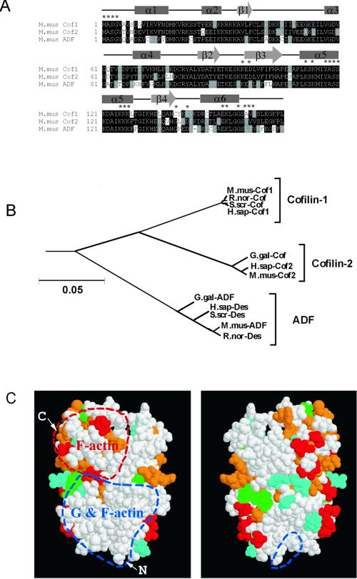
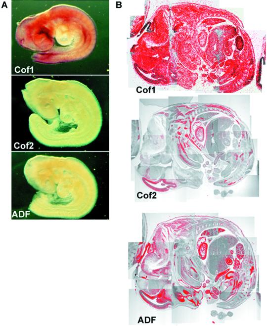
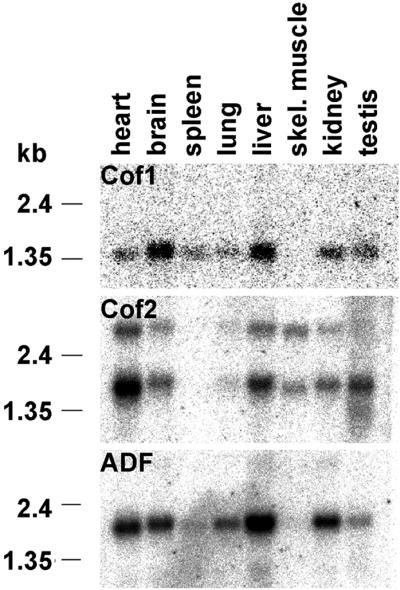
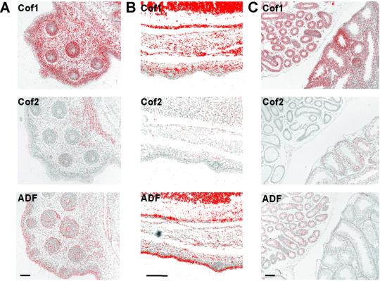
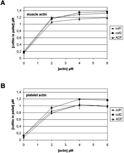
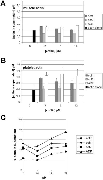
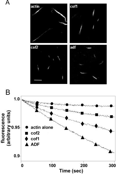
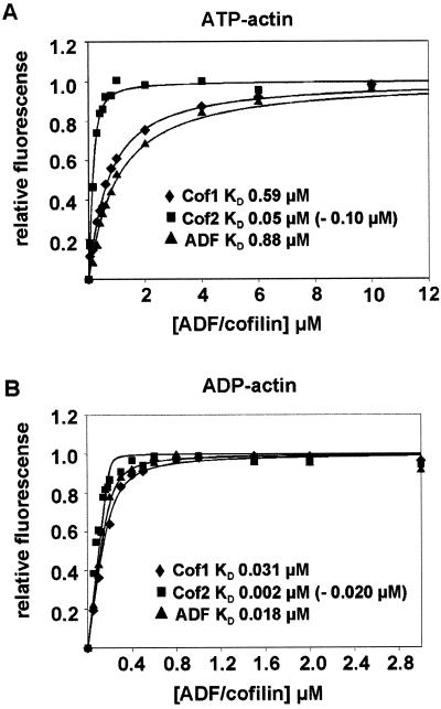
Actin-depolymerizing factor (ADF)/cofilins are essential regulators of actin filament turnover. Several ADF/cofilin isoforms are found in multicellular organisms, but their biological differences have remained unclear. Herein, we show that three ADF/cofilins exist in mouse and most likely in all other mammalian species. Northern blot and in situ hybridization analyses demonstrate that cofilin-1 is expressed in most cell types of embryos and adult mice. Cofilin-2 is expressed in muscle cells and ADF is restricted to epithelia and endothelia. Although the three mouse ADF/cofilins do not show actin isoform specificity, they all depolymerize platelet actin filaments more efficiently than muscle actin. Furthermore, these ADF/cofilins are biochemically different. The epithelial-specific ADF is the most efficient in turning over actin filaments and promotes a stronger pH-dependent actin filament disassembly than the two other isoforms. The muscle-specific cofilin-2 has a weaker actin filament depolymerization activity and displays a 5-10-fold higher affinity for ATP-actin monomers than cofilin-1 and ADF. In steady-state assays, cofilin-2 also promotes filament assembly rather than disassembly. Taken together, these data suggest that the three biochemically distinct mammalian ADF/cofilin isoforms evolved to fulfill specific requirements for actin filament dynamics in different cell types.


Figures

Figure 1
Figure 1
Comparison of the three mouse ADF/cofilin sequences. (A) Sequence alignment of the three mouse ADF/cofilins. The positions of the secondary structure elements based on the nuclear magnetic resonance structure of human destrin (Hatanaka et al., 1996) are indicated with boxes (α-helixes) and arrows (β-sheets). Asterisks above the sequences indicate residues that have been implicated in actin binding (Lappalainen et al. 1997; Ono et al. 1999, 2001; Van Troys et al. 2000). (B) Phylogenetic tree of all known mammalian and avian ADF/cofilins. The neighbor joining tree was produced by the Clustal-X software. A bar showing 5% divergence is included. Protein names, database, and accession numbers for the sequences, respectively, are listed below. Mus musculus cofilin-1: Swiss-Prot, P18760, Rattus norvegicus cofilin, Swiss-Prot, P45592, S. scrofa cofilin: GenBank, M20866, Homo sapiens cofilin-1: EMBL, X95404, Gallus gallus cofilin: GenBank, M55659, H. sapiens cofilin-2: EMBL, AF134802, M. musculus cofilin-2: Swiss-Prot, P45591, G. gallus ADF: GenBank, J02912, H. sapiens destrin: PIR, A54184, S. scrofa destrin: DDBJ, D90053, M. musculus ADF: EMBL, AB025406, R. norvegicus destrin: PIR, JE0223. (C) Space-filling model of human destrin (Hatanaka et al., 1996) in two different orientations (rotated 180o horizontally). Residues that correspond to variable amino acids between all three ADF/cofilins are indicated in red. The variable residues specific for cofilin-1, cofilin-2, and ADF are indicated in green, cyan, and orange, respectively. Regions that participate in actin monomer- and filament binding, and those involved only in actin filament binding are circled by red and blue dashed lines, respectively. Arrows mark the amino and carboxy terminus of the protein.
Figure 2
Figure 2
Expression of ADF/cofilins during development. (A) Whole mount in situ hybridization of E9.5 mouse embryos. Cofilin-1 mRNA (top) is expressed throughout the embryo, whereas only very weak cofilin-2 (middle) or ADF (bottom) expression can be detected under the same conditions. (B) In situ hybridization of E14 mouse embryo section. Cofilin-1 mRNA (top) shows ubiquitous expression. Expression of cofilin-2 (middle) is detected in developing muscles, such as in the tongue and tail. ADF (bottom) is expressed in developing epithelial cells, e.g., in intestine and skin.
Figure 3
Figure 3
Northern blot analyses of ADF/cofilin expression in adult mice. Cofilin-1 mRNA (top) is expressed at high levels in brain and liver; at moderate amounts in heart, spleen, lung, kidney, and testis; but is not expressed in skeletal muscle. The only isoform expressed in skeletal muscle is cofilin-2 (middle), which is also expressed at moderate levels in heart, liver, and testis, and at lower amounts in other organs. ADF (bottom) is expressed strongly in liver and at moderate levels in other organs, but is not found in skeletal muscle.
Figure 4
Figure 4
Expression of ADF/cofilins in different tissues. (A) In situ hybridization of an E15 mouse embryo's whisker pad. Cofilin-1 mRNA (top) is expressed all cell types of the whisker pad, whereas cofilin-2's (middle) expression is confined to the muscles behind the pad. ADF (bottom) is expressed in the epidermis and in the epithelia of whisker hair follicles. (B) In situ hybridization of E16 mouse embryo's back skin. Cofilin-1 (top) is found throughout the skin, whereas ADF (bottom) is concentrated especially to the outermost layers of the epidermis. Cofilin-2 (middle) is not expressed in the skin. (C) In situ hybridization of adult mouse testes. Cofilin-1 (top) is expressed in the testes and the epididymides. Cofilin-2 (middle) is expressed in the testes, but not in the epididymides. On the contrary, ADF (bottom) is expressed in the epididymal epithelia but not in the testis. Bar, 100 μm.
Figure 5
Figure 5
Binding of mouse ADF/cofilins to actin filaments. ADF/cofilins (1.5 μM) were mixed with 0, 2, 4, or 6 μM muscle (A) or platelet (B) actin at pH 7.5 and actin filaments were sedimented by centrifugation. The amount of ADF/cofilins in the pellet fraction was quantified from three independent experiments. All ADF/cofilins bound equally well to muscle and platelet actin filaments. The small differences in the levels of cosedimented ADF/cofilins result from the differences in their ability to promote actin filament disassembly (Figure 6).
Figure 6
Figure 6
Ability of ADF/cofilins to shift actin to the monomeric fraction in actin filament sedimentation assay. Muscle (3 μM) (A) or platelet actin (B) were mixed with 0, 3, 6, or 12 μM ADF/cofilins, actin filaments were sedimented by centrifugation, and the amount of actin in the supernatant and pellet fractions was quantified from three independent experiments. This assay was carried out at pH 7.5. Both cofilin-1 and ADF promote significant increases in the amount of monomeric actin and the ability of ADF to disassemble actin filaments is more pronounced with higher protein concentrations. In contrast to cofilin-1 and ADF, cofilin-2 is not able to promote actin filament disassembly. ADF/cofilins shifted more platelet actin (B) to the monomeric pool than muscle actin (A). (C) pH dependency of actin filament disassembly by mouse ADF/cofilins was studied with 3 μM platelet actin and 3 μM ADF/cofilins at pH 7.0–8.5. The ability of cofilin-1 and cofilin-2 to increase the amount of actin monomers is not greatly affected by pH. In contrast, the ability of ADF to disassemble actin filaments is significantly increased at higher pH.
Figure 7
Figure 7
Depolymerization/fragmentation of actin filaments by ADF/cofilins. (A) Labeled Alexa 488-actin (50%) was polymerized at pH 8.0 and mixed with ADF/cofilins to yield a final concentration of 2 μM for both proteins. Samples were visualized ∼40 s after mixing the two proteins. All three mouse ADF/cofilins shortened filaments, suggesting that they depolymerized/fragmented actin filaments in this assay. Bar 5, μM. (B) Turnover of actin filaments in the presence and absence of ADF/cofilins was measured by following the release of ε-ATP from the filaments at pH 8.0 after an ATP chase. The final concentrations of actin and ADF/cofilins in this assay were 17 and 4.3 μM, respectively. The decrease in the fluorescence of F-actin–bound ε-ATP represents the turnover of actin filaments.
Figure 8
Figure 8
Interaction of ADF/cofilins with actin monomers. The increase in fluorescence of 0.2 μM NBD-labeled MgATP-G-actin (A) or Mg-ADP-G-actin (B) was measured at different concentrations of ADF/cofilins under physiological ionic conditions at pH 8.0. Symbols are data and lines are calculated binding curves. Each data point on the graph is a mean value of three independent experiments. Dissociation constants (KD) derived from the binding curves are indicated in the figure. The two values given for cofilin-2 result from two independent methods that were applied for determination of the concentration of this protein.

References

    1. Abe H, Endo T, Yamamoto K, Obinata T. Sequence of cDNAs encoding actin depolymerizing factor and cofilin of embryonic chicken skeletal muscle: two functionally distinct actin-regulatory proteins exhibit high structural homology. Biochemistry. 1990;29:7420–7425. - PubMed
    1. Adams ME, Minamide LS, Duester G, Bamburg JR. Nucleotide sequence and expression of a cDNA encoding chick brain actin depolymerizing factor. Biochemistry. 1990;29:7414–7420. - PubMed
    1. Ayscough KR. In vivo functions of actin-binding proteins. Curr Opin Cell Biol. 1998;10:102–111. - PubMed
    1. Bamburg JR. Proteins of the ADF/cofilin family: essential regulators of actin dynamics. Annu Rev Cell Dev Biol. 1999;15:185–230. - PubMed
    1. Bamburg JR, Bray D. Distribution and cellular localization of actin depolymerizing factor. J Cell Biol. 1987;105:2817–2825. - PMC - PubMed

Publication types

MeSH terms