Comparative Study
. 2002 Jan;13(1):195-210.
doi: 10.1091/mbc.01-05-0221.

Evidence that ternary complex (eIF2-GTP-tRNA(i)(Met))-deficient preinitiation complexes are core constituents of mammalian stress granules

Affiliations
Comparative Study

Evidence that ternary complex (eIF2-GTP-tRNA(i)(Met))-deficient preinitiation complexes are core constituents of mammalian stress granules

Nancy Kedersha et al. Mol Biol Cell. 2002 Jan.

Abstract

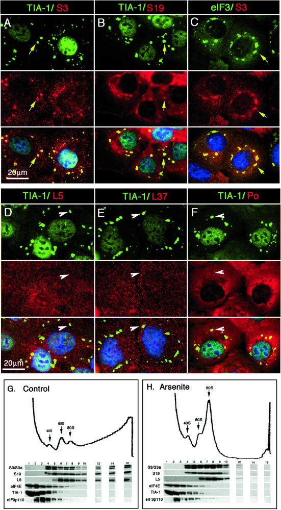
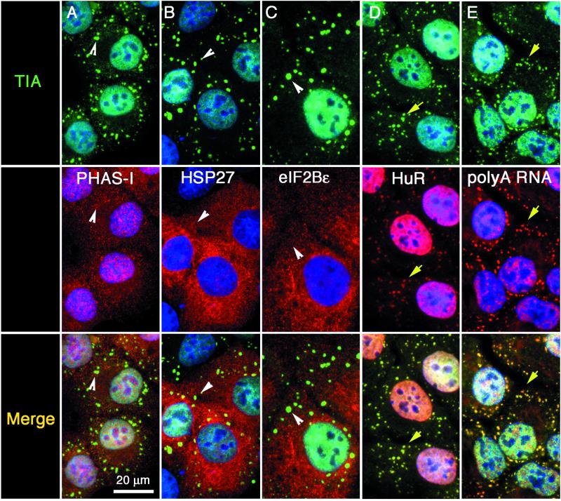
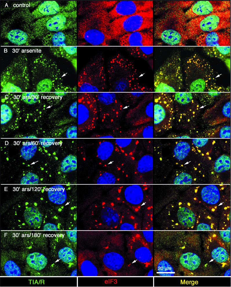
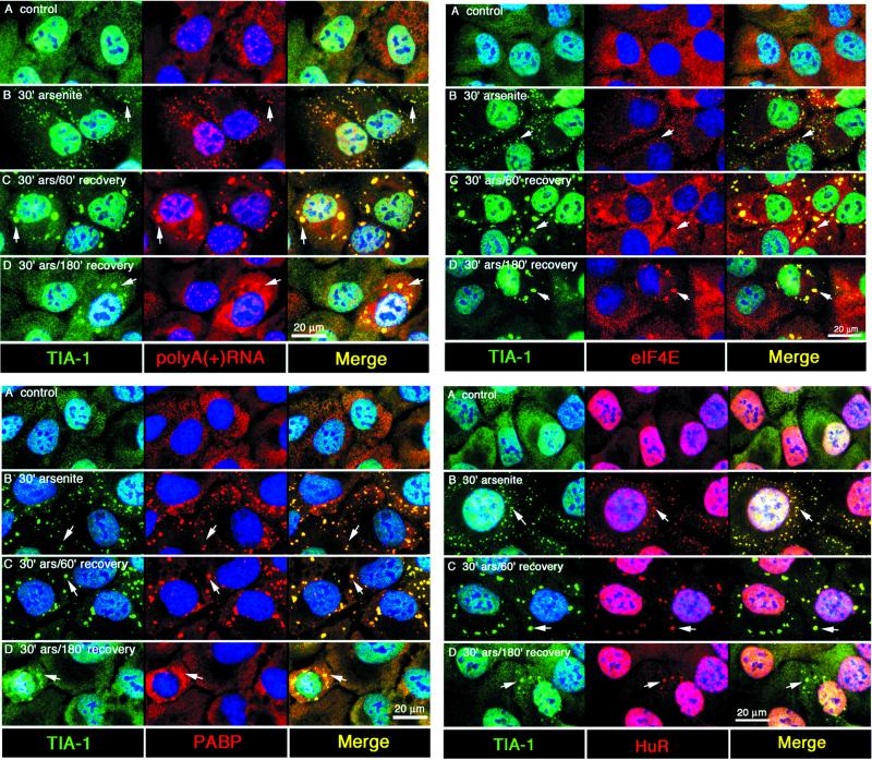
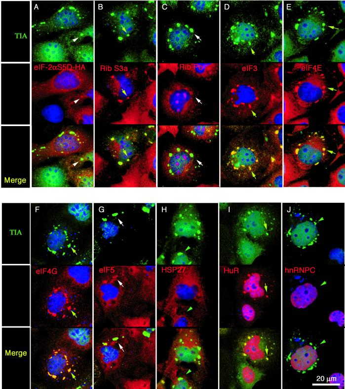
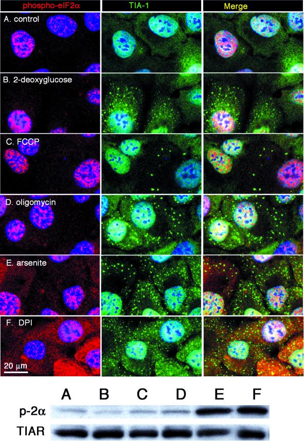
Environmental stress-induced phosphorylation of eIF2alpha inhibits protein translation by reducing the availability of eIF2-GTP-tRNA(i)Met, the ternary complex that joins initiator tRNA(Met) to the 43S preinitiation complex. The resulting untranslated mRNA is dynamically routed to discrete cytoplasmic foci known as stress granules (SGs), a process requiring the related RNA-binding proteins TIA-1 and TIAR. SGs appear to be in equilibrium with polysomes, but the nature of this relationship is obscure. We now show that most components of the 48S preinitiation complex (i.e., small, but not large, ribosomal subunits, eIF3, eIF4E, eIF4G) are coordinately recruited to SGs in arsenite-stressed cells. In contrast, eIF2 is not a component of newly assembled SGs. Cells expressing a phosphomimetic mutant (S51D) of eIF2alpha assemble SGs of similar composition, confirming that the recruitment of these factors is a direct consequence of blocked translational initiation and not due to other effects of arsenite. Surprisingly, phospho-eIF2alpha is recruited to SGs that are disassembling in cells recovering from arsenite-induced stress. We discuss these results in the context of a translational checkpoint model wherein TIA and eIF2 are functional antagonists of translational initiation, and in which lack of ternary complex drives SG assembly.


Figures

Figure 1
Figure 1
Small, but not large ribosomal subunits, are recruited to SGs. DU145 cells were exposed to 1 mM sodium arsenite for 30 min and allowed to recover for 2 h to generate SGs of maximal size. Cells were fixed and stained for TIA-1 to reveal SGs (green, panels A, B, D, E, and F), and counterstained with affinity-purified antibodies against different ribosomal proteins: ribosomal protein S3/3a (A and C, red), ribosomal protein S19 (B, red), ribosomal protein L5 (D, red), and ribosomal protein L37 (E, red). (F) Localization of ribosome Po proteins (red) detected using a human auto-antisera. Affinity-purified antisera against eIF3 was used to reveal the colocalization of eIF3 (C, green) with SG-associated ribosomal S3/3a (C, red). Yellow arrows indicate recruitment of ribosomal proteins to SGs; white arrowheads indicate SGs to which large ribosomal subunit proteins are not recruited. Bar, 20 μm. Sedimentation of S20 extracts of control (G) versus arsenite-treated (H) DU145 cells were resolved on 10–47% sucrose gradients, eluted from the top, and the elution profile at OD254 was recorded. Fractions were collected and subjected to SDS-PAGE and Western blotting by using antibodies to components of the small (e.g., S3/S3a and S19) or large (e.g., L5, L37, and Po) ribosomal subunit, as well as the eIF4 component eIF4E, TIA-1, and eIF3.
Figure 2
Figure 2
Components of the 48S preinitiation complex are recruited to stress granules. DU145 cells were treated with arsenite (0.5 mM, 1 h) and processed for multicolor immunofluorescence microscopy by using antibodies reactive with TIA-1/R (green), the indicated initiation factor or other protein (red), and DNA (blue). Each vertical column shows views of the same field. Top row: TIA-1 or TIAR (green). Middle row (red): eIF2α (A), eIF3 (B), eIF4E (C), eIF4G (D), and eIF5 (E). Bottom row, merged views. Yellow arrows point out individual stress granules in which both the red and the green signals overlap; white arrowheads indicate the position of SGs that only contain TIA. Nuclei are stained blue. Bar, 20 μm.
Figure 3
Figure 3
Other factors present or absent from stress granules. DU145 cells were stimulated with arsenite as described in Figure 2 and processed for immunofluorescence. Each vertical column shows views of the same field. Top row: TIA-1 or TIAR (green). Middle row (red): PHAS-I (eIF4E-binding protein) (A), HSP27 (B), eIF2Bε (C), and HuR (D); poly(A)+ RNA (E). Bottom row, merged views. Yellow arrows point out individual stress granules in which both the red and the green signals overlap; white arrowheads indicate the position of SGs that only contain TIA. Nuclei are stained blue. Bar, 20 μm.
Figure 4
Figure 4
Recombinant HA-eIF2α, and HA-eIF2β are not recruited to SGs; recombinant HA-eIF3p48 is recruited to SGs. COS cells were transiently transfected with HA-eIF2α (A and D; green), HA-eIF2β (B and E; green); or HA-eIF3p48/int6 (C and F; green). Untreated cells (A–C) or arsenite-treated cells (D–F) were fixed and stained for HA (green, all panels) and TIA-1 (red, all panels). Arrows indicate SGs in which the HA-tagged protein is recruited to the SG; arrowheads indicate SGs where no recruitment of the HA-tagged protein is detected. Bar, 20 μm.
Figure 5
Figure 5
Kinetics of the colocalization of TIAR and eIF3 at stress granules. DU145 cells were treated with 1.0 mM sodium arsenite for 30 min, washed, and then further incubated in the absence of arsenite (recovery) for the indicated times. Each row depicts parallel views of the same field. TIA-1/R appears green, eIF3 appears red, and coincidence appears yellow (right column: merge). Control (A), 30-min arsenite treatment without recovery (B), 30-min arsenite followed by 30-min recovery (C); 30-min arsenite followed by 1-h recovery (D); 30-min arsenite followed by 2-h recovery (E); and 30-min arsenite followed by 3-h recovery (F). Nuclei are stained blue. Bar, 20 μm.
Figure 6
Figure 6
Coordinate recruitment of poly(A)+ RNA, PABP-I, eIF4E, and HuR to SGs during SG assembly and disassembly. DU145 cells were arsenite-stressed and allowed to recover as in Figure 5. Top left, TIA-1 (green) versus poly(A) (red); bottom left, TIA-1 (green), PABP (red); Top right, TIA-1 (green), eIF4E (red); bottom right, TIA-1(green), HuR (red). In all panels, blue represents DNA (Hoechst staining). Bar, 20 μm. Merged views are shown in the right panels of each series. Arrows indicate SGs in each view.
Figure 7
Figure 7
Composition of eIF2α (S51D)-induced stress granules. COS cells were transiently transfected with a plasmid encoding recombinant phosphomimetic eIF2α (S51D) to induce the assembly of stress granules. After 28 h, cells were processed for two-color immunofluorescence microscopy by using antibodies reactive with TIA-1 or TIAR (green) and the indicated translation factors (red). Each column contains paired views of the same field, with the merged image at the bottom. Shown in red are HA-eIF2α (S51D) (A); small ribosomal protein S3a (B); large ribosomal subunit protein Po (C); eIF3 (D); eIF4E (E); eIF4G (F); eIF5 (G); HSP27 (H); HuR (I); and hnRNP C (J). SGs appear yellow/orange when the indicated translation factor colocalizes with TIA-1 (eIF3, eIF4E, eIF4G, ribosomal subunit S3, HuR; yellow arrows) or green when the indicated translation factor does not colocalize with TIA-1 (e.g., HSP27, hnRNP C; green arrowheads). Minimal colocalization is seen with HA-eIF2α (S51D), ribosome P antigen, and eIF-5 (white arrows). Nuclei are stained blue. Bar, 20 μm.
Figure 8
Figure 8
Phospho-eIF2α is only present in stress granules during recovery. DU145 cells were treated with arsenite and allowed to recover as described in Figure 5 and then stained for TIA-1 and phospho-eIF2α. Left column, TIA-1 (green) and DNA (blue); middle column, phospho-eIF2α(red) and DNA (blue); right column, merge. Control (A), 30-min arsenite treatment without recovery (B), 30-min arsenite followed by 30-min recovery (C), 30-min arsenite followed by 1-h recovery (D), 30-min arsenite followed by 2-h recovery (E), and 30-min arsenite followed by 3-h recovery (F). Bars, 20 μm. (G) Immunoblot of equal amounts of total protein lysates from cells treated identically as described above, probed with phospho-eIF2α antibody. (H) Phospho-mimetic HA-eIF2αS51D was expressed in COS and detected with anti-HA (green). Cells were counterstained for TIA-1 (red) and DNA (blue). Arrows indicate position of SG, to which a minor but detectable fraction of the HA-tagged protein is localized. Bar, 20 μm.
Figure 9
Figure 9
Induction of SGs by energy depletion without inducing eIF2α phosphorylation. DU145 cells were treated with 2-deoxyglucose (B), FCCP (C), oligomycin (D), arsenite (E), or DPI (F) in either glucose-free media (B, C, D, and F) or regular media (E) for 30 min. Cells were then fixed and stained or lysed directly in SDS-buffer for PAGE. (Top) Immunofluorescence showing TIA-1 in green and phospho-eIF2α in red. Nuclei are stained blue. Bars, 20 μm. (Bottom) Immunoblot of total cell lysates treated as described above, probed with phospho-eIF2α and TIAR (as loading control).

References

    1. Berlanga J, Herrero S, DeHaro C. Characterization of the hemin-sensitive eukaryotic initiation factor 2a kinase from mouse nonerythroid cells. J Cell Biol. 1998;273:32340–32346. - PubMed
    1. Bernstam L, Nriagu J. Molecular aspects of arsenic stress. J Toxicol Environ Health B Crit Rev. 2000;3:293–322. - PubMed
    1. Cuesta R, Laroia G, Schneider RJ. Chaperone Hsp27 inhibits translation during heat shock by binding eIF4G and facilitating dissociation of cap-initiation complexes. Genes Dev. 2000;14:1460–1470. - PMC - PubMed
    1. Del Gatto-Konczak F, Bourgeois CF, Le Guiner C, Kister L, Gesnel MC, Stevenin J, Breathnach R. The RNA-binding protein TIA-1 is a novel mammalian splicing regulator acting through intron sequences adjacent to a 5′ splice site. Mol Cell Biol. 2000;20:6287–6299. - PMC - PubMed
    1. Dever TE. Translation initiation: adept at adapting. Trends Biochem Sci. 1999;24:398–403. - PubMed

Publication types

MeSH terms

LinkOut - more resources