Comparative Study
. 2002 Jan;13(1):225-37.
doi: 10.1091/mbc.01-07-0376.

Absence of direct delivery for single transmembrane apical proteins or their "Secretory" forms in polarized hepatic cells

Affiliations
Comparative Study

Absence of direct delivery for single transmembrane apical proteins or their "Secretory" forms in polarized hepatic cells

M Bastaki et al. Mol Biol Cell. 2002 Jan.

Abstract

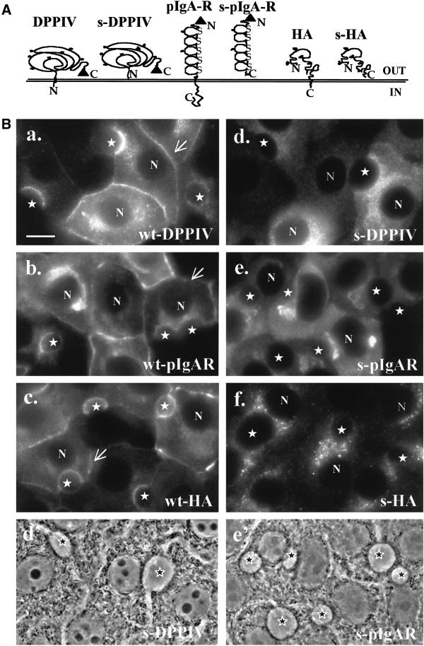
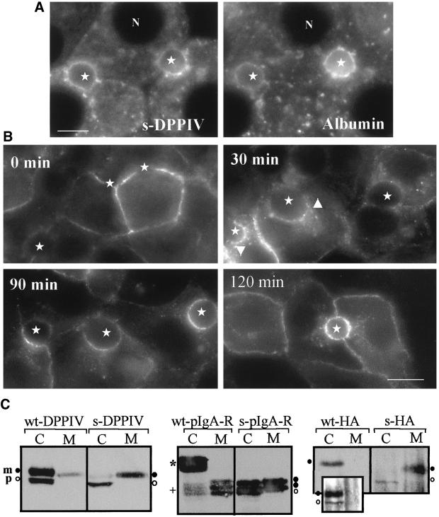
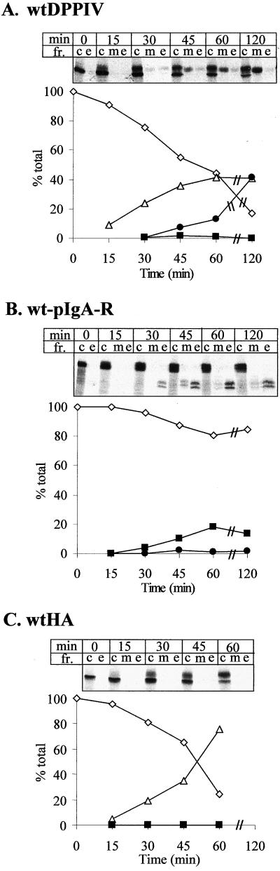
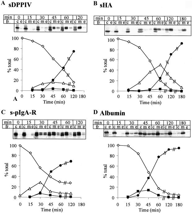
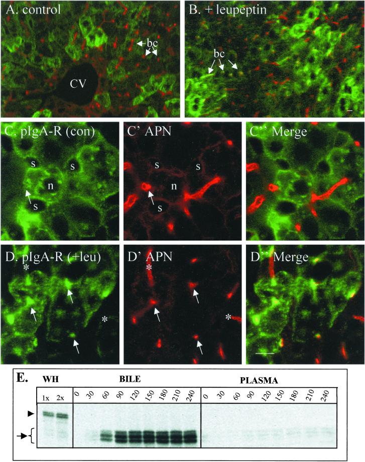
The absence of a direct route to the apical plasma membrane (PM) for single transmembrane domain (TMD) proteins in polarized hepatic cells has been inferred but never directly demonstrated. The genes encoding three pairs of apical PM proteins, whose extracellular domains are targeted exclusively to the apical milieu in Madin-Darby canine kidney cells, were packaged into recombinant adenovirus and delivered to WIF-B cells in vitro and liver hepatocytes in vivo. By immunofluorescence and pulse-chase metabolic labeling, we found that the soluble constructs were overwhelmingly secreted into the basolateral milieu, which in vivo is the blood and in vitro is the culture medium. The full-length proteins were first delivered to the basolateral surface but then concentrated in the apical PM. Our results imply that hepatic cells lack trans-Golgi network (TGN)-based machinery for directly sorting single transmembrane domain apical proteins and raise interesting questions about current models of PM protein sorting in polarized and nonpolarized cells.


Figures

Figure 1
Figure 1
Expression of the wt- and s-forms of DPPIV, pIgA-R, and HA in WIF-B cells. (A) Constructs used in the study. ▴, epitope tags used; s=secretory; N, aminoterminus; C, carboxy terminus. (B) Cells were infected with one of the indicated viruses and fixed 24 h later. The expressed proteins were visualized by indirect immunofluorescence. The wt-proteins (a–c) concentrate at the apical PM (stars) but are also at the basolateral PM (arrows) and in the perinuclear area (N, nucleus). The secretory forms (d–f) are detected only intracellularly and not in the BC space (stars). Phase-contrast images of infected WIF-B cells (e, s-DPPIV; f, s-pIgAR) are shown. Bar, 10 μm.
Figure 2
Figure 2
Behavior of wt- and s-recombinant proteins in WIF-B cells. (A) Dispersal. Infected WIF-B cells treated with nocodazole (33 μM) for 2 h before fixation were double-labeled with α-V5 to detect s-DPPIV (left) and α-rat serum albumin (right). The Golgi is dispersed and the s-proteins ring the BC (stars). N, nucleus. Bar, 10 μm. (B) Transcytosis. HA expressed at the basolateral surface was labeled with α-HA (R584, 4°C for 15 min) and chased at 37°C for the indicated times before fixation and visualization by secondary antibody. Note the absence of labeling at the apical PM after 0 min of chase (stars) and the progressive increase with longer times at 37°C. White arrowheads point to puncta near the apical PM. Bar, 10 μm. (C) Maturation and secretion. Cells were infected with the indicated virus and 24 h later, cell extracts (100%) and conditioned media (20%) were immunoprecipitated and processed for immunoblot analysis. C, cell; M, medium; ●, mature (m); ○, precursor (p); *, full-length pIgA-R; +, truncated p-IgA-R.
Figure 3
Figure 3
Quantitative analysis of recombinant wt-protein expression in WIF-B cells. WIF-B cells infected with the indicated adenovirus were pulse-labeled with 35S-amino acids for 5 min and chased for up to 120 min. Samples were collected at 15-min intervals for the first hour and each hour thereafter. The 35S-proteins were immunoprecipitated from equal portions of the collected samples and processed for autoradiography. fr., fraction; c, cell extracts; m, medium (basolateral secretion); e, EGTA extract (apical secretion). Each value in the graphs is expressed as a percentage of the total (c + m + e = 100%) at each time point. ⋄, precursor; ▵, mature; ●, medium; ▪, EGTA extract. Values are averages of duplicates from the same experiment as shown in the gels. Two experiments gave similar results.
Figure 4
Figure 4
Quantitative analysis of recombinant s-protein expression in WIF-B cells. WIF-B cells infected with the indicated adenovirus were pulse-labeled with 35S-amino acids for 5 min and chased up to 180 min. 35S-labeled samples were processed as described in Figure 7 legend. D: Endogenous 35S-albumin was also immunoprecipitated from the same samples in some of these experiments. Symbols are described in Figure 7 legend. Two experiments gave similar results.
Figure 5
Figure 5
Recombinant wt-pIgA-R expressed in vivo reaches the apical surface of hepatocytes and is cleaved. Mice injected with V5-wt-pIgA-R adenovirus were sacrificed 3 d later, without (A and B, control) or with (C and D, +leupeptin) ip injection of 1 mg/100 gm body weight leupeptin administered every 30 min for 4 h. 7 um fixed sections were labeled with mouse α-V5-FITC (green) and rabbit α-APN then Alexa- α-rabbit antibody (red). Cells labeled with the α-V5 are those infected. In panel A, there is no overlap between the red and green signals, indicating the absence of pIgA-R at the apical PM of infected cells. However, in panel B, from an animal treated with leupeptin, recombinant wt-pIgA-R is seen at the apical PM and overlaps with endogenous APN. (bc, bile canaliculi). Rows C and D are enlarged views of untreated (C) and leupeptin-treated (D) livers. Symbols: n, nuclei; s, sinusoidal space; arrows, bile canaliculi; asterisks in D and D', bile canalicular PM in uninfected cells. Bars, 20 um (A and B) and 10 um (C and D). E. Rats infected with adenovirus were pulse-chased with 35S-cysteine. 35S-pIgA-R was immunoprecipitated with α-V5 from WHs, bile and plasma collected at the indicated times. 15% of each bile sample and 0.05% of each plasma sample are represented in the individual lanes in the autoradiogram. Symbols: arrowhead, mature forms of wt-pIgA-R; arrow, cleaved forms.

References

    1. Aroeti B, Okhrimenko H, Reich V, Orzech E. Polarized trafficking of plasma membrane proteins: emerging roles for coats, SNAREs, GTPases and their link to the cytoskeleton. Biochim Biophys Acta. 1998;1376:57–90. - PubMed
    1. Barr VA, Hubbard AL. Newly synthesized hepatocyte plasma membrane proteins are transported in transcytotic vesicles in the bile duct-ligated rat. Gastroenterology. 1993;105:554–571. - PubMed
    1. Bartles JR, Feracci HM, Stieger B, Hubbard AL. Biogenesis of the rat hepatocyte plasma membrane in vivo: comparison of the pathways taken by apical and basolateral proteins using subcellular fractionation. J Cell Biol. 1987;105:1241–1251. - PMC - PubMed
    1. Brown DA, London E. Functions of lipid rafts in biological membranes. Annu Rev Cell Dev Biol. 1998;14:111–136. - PubMed
    1. Casanova JE, Hubbard AL, Mishumi Y, Ikehara Y, Mostov KE. Direct apical sorting of rat liver dipeptidylpeptidase IV expressed in MDCK cells. J Biol Chem. 1991;266:24428–24432. - PubMed

Publication types

MeSH terms

LinkOut - more resources