Comparative Study
. 2002 Jan;13(1):348-61.
doi: 10.1091/mbc.01-05-0225.

Cell proliferation and DNA breaks are involved in ultraviolet light-induced apoptosis in nucleotide excision repair-deficient Chinese hamster cells

Affiliations
Comparative Study

Cell proliferation and DNA breaks are involved in ultraviolet light-induced apoptosis in nucleotide excision repair-deficient Chinese hamster cells

Torsten R Dunkern et al. Mol Biol Cell. 2002 Jan.

Abstract

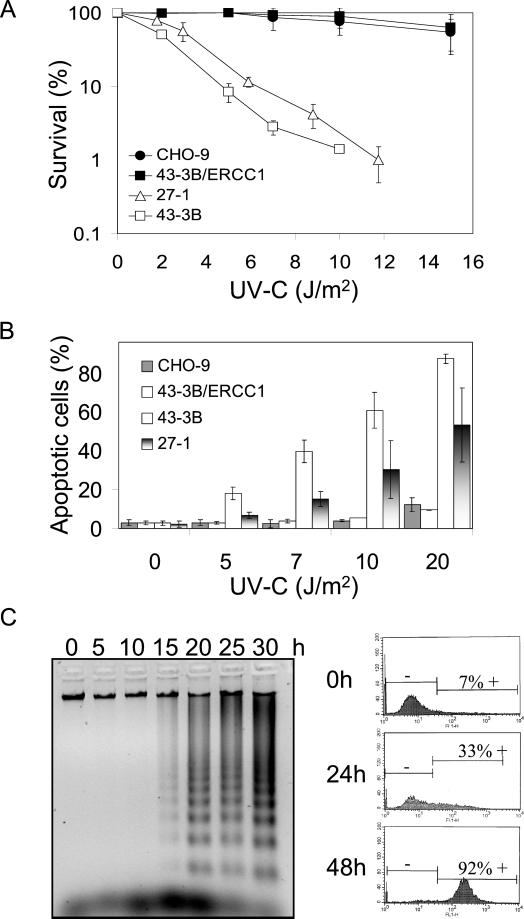
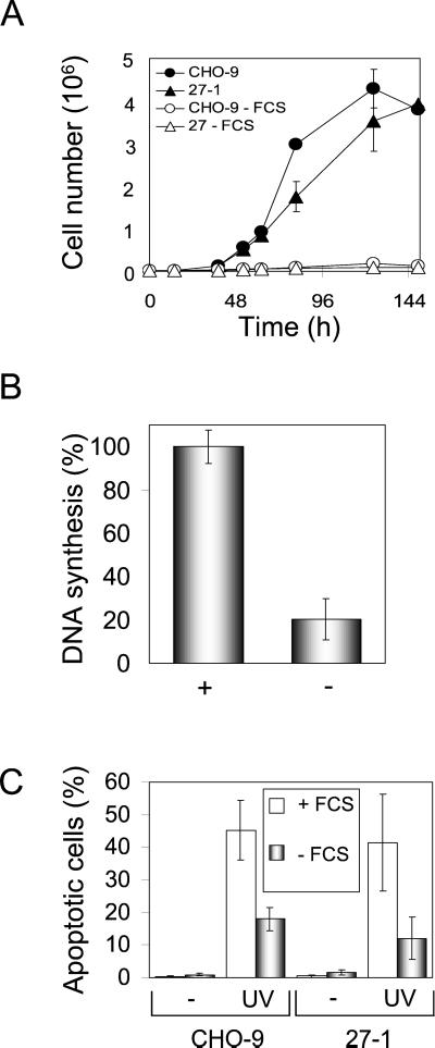
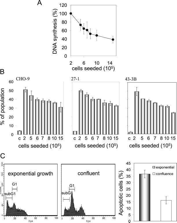
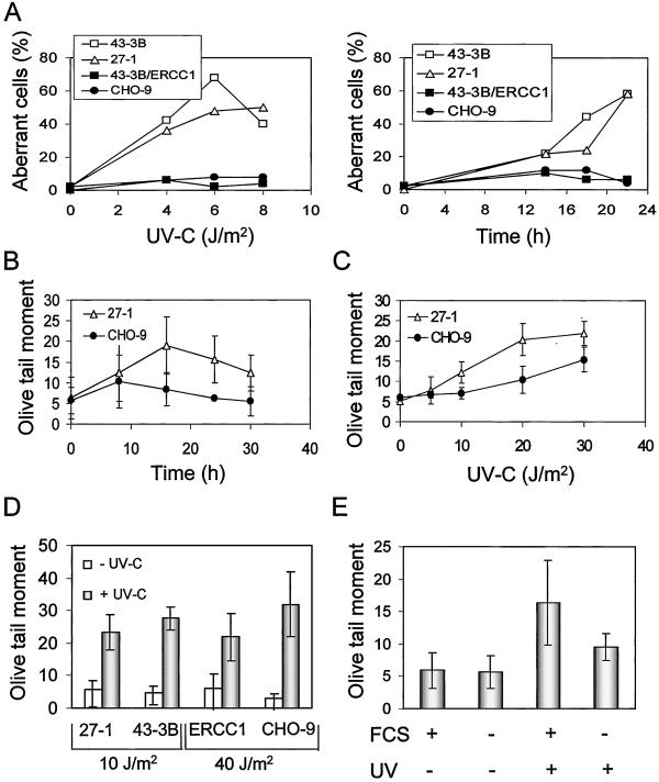
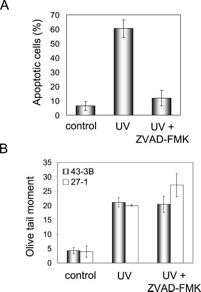
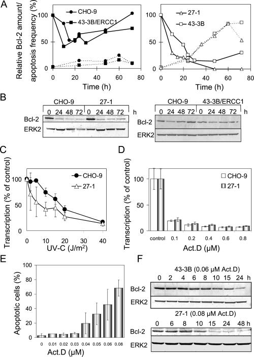
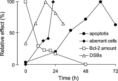
UV light targets both membrane receptors and nuclear DNA, thus evoking signals triggering apoptosis. Although receptor-mediated apoptosis has been extensively investigated, the role of DNA damage in apoptosis is less clear. To analyze the importance of DNA damage induced by UV-C light in apoptosis, we compared nucleotide excision repair (NER)-deficient Chinese hamster ovary cells (lines 27-1 and 43-3B mutated for the repair genes ERCC3 and ERCC1, respectively) with the corresponding DNA repair-proficient fibroblasts (CHO-9 and ERCC1 complemented 43-3B cells). NER-deficient cells were hypersensitive as to the induction of apoptosis, indicating that apoptosis induced by UV-C light is due to unrepaired DNA base damage. Unrepaired lesions, however, do not activate the apoptotic pathway directly because apoptosis upon UV-C irradiation requires DNA replication and cell proliferation. It is also shown that in NER-deficient cells unrepaired lesions are converted into DNA double-strand breaks (DSBs) and chromosomal aberrations by a replication-dependent process that precedes apoptosis. We therefore propose that DSBs arising from replication of DNA containing nonrepaired lesions act as an ultimate trigger of UV-C-induced apoptosis. Induction of apoptosis by UV-C light was related to decline in the expression level of Bcl-2 and activation of caspases. Decline of Bcl-2 and subsequent apoptosis might also be caused, at least in part, by UV-C-induced blockage of transcription, which was more pronounced in NER-deficient than in wild-type cells. This is in line with experiments with actinomycin D, which provoked Bcl-2 decline and apoptosis. UV-C-induced apoptosis due to nonrepaired DNA lesions, replication-dependent formation of DSBs, and activation of the mitochondrial damage pathway is independent of functional p53 for which the cells are mutated.


Figures

Figure 1
Figure 1
Cell-killing response and apoptosis in NER-deficient cells treated with UV-C light. (A) Survival of CHO-9, 43-3B/ERCC1, 27-1, and 43-3B cells as a function of UV-C dose, as determined by colony formation assay. (B) Frequency of apoptosis of CHO-9, 43-3B/ERCC1, 43-3B, and 27-1 cells as a function of dose of UV-C (measured 48 h after irradiation). (C) Apoptotic DNA laddering pattern in 27-1 cells (left) as a function of time after UV-C treatment (10 J/m2). Flow cytometric quantification of 27-1 cells with enhanced caspase activity 24 and 48 h after UV-C irradiation (10 J/m2) as determined by FITC-zVAD-FMK staining (right).
Figure 2
Figure 2
UV-C–induced apoptosis in serum-starved cells. (A) Growth curves of CHO-9 and 27-1 cells cultured with or without fetal calf serum. (B) DNA synthesis of CHO cells cultured for 72 h with (+) or without (−) serum as determined by BrdU-ELISA. (C) Frequency of apoptosis in CHO-9 and 27-1 cells irradiated with 40 and 10 J/m2, respectively, as measured by annexin staining. Cells were cultured for 72 h with or without serum before irradiation and apoptosis was measured 48 h after irradiation.
Figure 3
Figure 3
Dependency of UV-C–induced apoptosis on proliferation. (A) DNA synthesis as a function of the number of cells seeded. (B) Frequency of apoptosis 48 h after UV-C irradiation (10 J/m2 for 27-1 and 43-3B cells; 40 J/m2 for CHO-9 cells) as a function of cell seeding number. (C) Flow cytometric quantification of UV-C (40 J/m2)-induced apoptotic sub-G1 cells in exponentially growing and confluent primary mouse fibroblasts as measured 48 h after irradiation.
Figure 4
Figure 4
UV-C–induced DNA breakage is proliferation dependent. (A) Frequency of UV-C–induced chromosomal aberrations in 43-3B, 27-1, 43-3B/ERCC1, and CHO-9 cells as a function of UV-C dose (left; 22 h post-treatment) and time (right; 8 J/m2 of UV-C). (B) UV-C–induced DSBs as a function of time. Cells were irradiated with a UV-C dose of 10 J/m2 and analyzed by means of neutral SCGE. (C) DSBs as a function of UV-C dose 20 h post-treatment, as determined by neutral SCGE. (D) UV-C light-induced DSBs as measured 20 h after UV-C irradiation in 27-1, 43-3B, 43-3B/ERCC1 (ERCC1), and CHO-9 cells, which were determined by neutral SCGE assay. (E) UV-C–induced DSBs 20 h after UV-C treatment (10 J/m2) in 27-1 cells cultivated in medium not containing (−) or containing (+) fetal calf serum, as determined by neutral SCGE.
Figure 5
Figure 5
Reduction of apoptosis but not DSBs after UV-C irradiation by zVAD-FMK. (A) Frequency of apoptosis in UV-C–irradiated (10 J/m2) 27-1 cells treated or not treated with the general caspase inhibitor zVAD-FMK (100 μM). Cells were analyzed 48 h after UV-C treatment by annexin staining and flow cytometry. (B) UV-C (10 J/m2) induced DSBs in 27-1 cells treated or not with the general caspase inhibitor zVAD-FMK (100 μM). Cells were harvested 20 h after irradiation and subjected to neutral SCGE.
Figure 6
Figure 6
Cell cycle-specific activation of caspases and p53 activity in UV-C–irradiated 27-1 cells. (A) Flow cytometric dual color staining for DNA content (PI) and caspase activity of untreated (top, left) and UV-C–treated (10 J/m2; top, right) 27-1 cells. The bottom panels show quantitative cell cycle distributions 36 h after treatment. (B) Determination of p53-regulated mdm2 promoter activity after transient transfection of the construct followed by UV-C irradiation (15 J/m2). Cells were harvested 12 h after treatment of BalbC, CHO-9, and 27-1 cells and analyzed for luciferase activity. (C) Expression of CD95R after UV-C treatment (10 J/m2) of 27-1 cells and of doxorubicin-treated (0.5 μg/ml) CHO-9 and BK4 cells, as shown by Western blot analysis.
Figure 7
Figure 7
Decline of Bcl-2 in NER-deficient cells after UV-C irradiation. (A) Relative Bcl-2 amount (continuous line) and apoptosis frequency (dotted line) as a function of time after UV-C irradiation (10 J/m2) of CHO-9 and 43-3B/ERCC1 cells (left) and NER-deficient 27-1 and 43-3B cells (right). (B) Expression of Bcl-2 in CHO-9 and 27-1 cells treated with 40 and 10 J/m2, respectively (left), as well as expression of Bcl-2 in 10 J/m2 UV-C–treated CHO-9 and 43-3B/ERCC1 cells, as shown by Western blot analysis. (C) Transcriptional activity as a function of UV-C dose in CHO-9 and 27-1 cells 4 h after treatment. (D) Transcriptional activity as a function of concentration of actinomycin D in CHO-9 and 27-1 cells, as determined 4 h after treatment. (E) Frequency of apoptosis in 27-1 cells as a function of concentration of actinomycin D. (F) Expression of Bcl-2 in 43-3B and 27-1 cells after actinomycin D treatment, as shown by Western blot analysis. The ERK2 signal illustrates equal protein loading on the gel.
Figure 8
Figure 8
Effect of Bcl-2 overexpression in 27-1 cells on UV-C–induced apoptosis. 27-1 cells were transfected with a myc-bcl-2 construct and seven Bcl-2-overexpressing clones were established. Clones, vector transfected cells, and untransfected cells were irradiated (10 J/m2 UV-C) and flow cytometric annexin measurements were performed 48 h later (∗∗∗, highly significant difference with p < 0.05). Ordinate: average yield (%) of apoptotic and necrotic cells.
Figure 9
Figure 9
Time course of effects induced by UV-C light in NER-deficient cells. Apoptosis, DSBs, chromosomal aberrations (percentage of aberrant cells) as well as Bcl-2 expression are shown at various time points after irradiation (10 J/m2) of 27-1 cells. Values are calculated as percentage relative to the maximum value.

References

    1. Agarwal ML, Agarwal A, Taylor WR, Stark GR. p53 controls both the G2/M and the G1 cell cycle checkpoints and mediates reversible growth arrest in human fibroblasts. Proc Natl Acad Sci USA. 1995;92:8493–8497. - PMC - PubMed
    1. Antonsson B, Martinou JC. The Bcl-2 protein family. Exp Cell Res. 2000;256:50–57. - PubMed
    1. Aragane Y, Kulms D, Metze D, Wilkes G, Pöppelmann B, Luger T, Schwarz T. Ultraviolet light induces apoptosis via direct activation of CD95 (Fas/APO-1) independently of its ligand CD95L. J Cell Biol. 1998;140:171–182. - PMC - PubMed
    1. Brash DE, Ziegler A, Jonason AS, Simon JA, Kunala S, Leffell DJ. Sunlight and sunburn in human skin cancer: p53, apoptosis, and tumor promotion. J Invest Dermatol Symp Proc. 1996;2:136–142. - PubMed
    1. Canman CE, Kastan MB. Signal transduction. Three paths to stress relief. Nature. 1996;384:213–214. - PubMed

Publication types

MeSH terms