. 2002 Feb 1;30(3):636-42.
doi: 10.1093/nar/30.3.636.

Action of prokaryotic enhancer over a distance does not require continued presence of promoter-bound sigma54 subunit

Affiliations

Action of prokaryotic enhancer over a distance does not require continued presence of promoter-bound sigma54 subunit

Vladimir Bondarenko et al. Nucleic Acids Res. .

Abstract

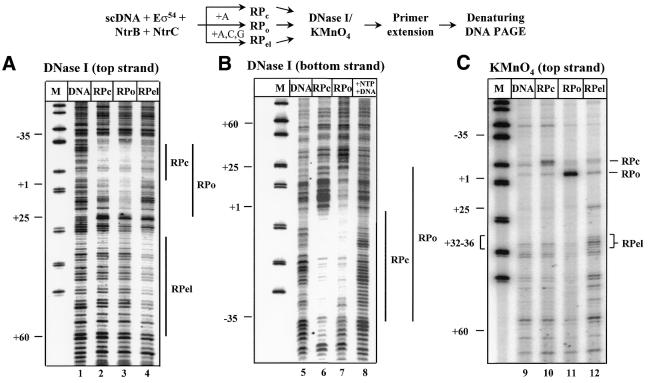
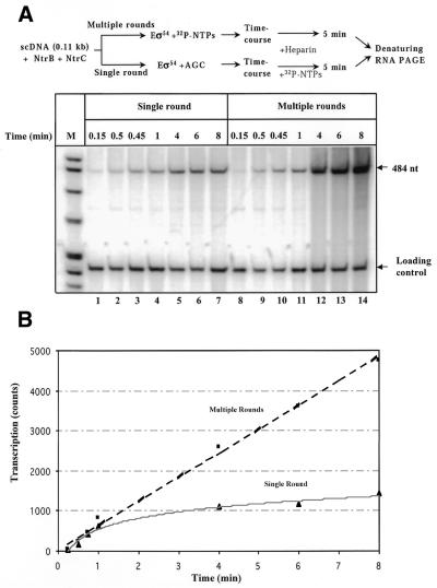
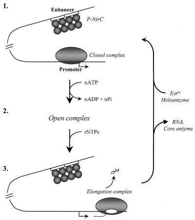
The mechanism by which an enhancer activates transcription over large distances has been investigated. Activation of the glnAp2 promoter by the NtrC-dependent enhancer in Escherichia coli was analyzed using a purified system supporting multiple-round transcription in vitro. Our results suggest that the enhancer-promoter interaction and the initiation complex must be formed de novo during every round of transcription. No protein remained bound to the promoter after RNA polymerase escaped into elongation. Furthermore, the rate of initiation during the first and subsequent rounds of transcription were very similar, suggesting that there was no functional 'memory' facilitating multiple rounds of transcription. These studies exclude the hypothesis that enhancer action during multiple-round transcription involves the memory of the initial activation event.


Figures

Figure 1
Figure 1
Templates for analysis of the mechanism of glnAp2 promoter activation by NtrC-dependent enhancer. Plasmid templates having different enhancer–promoter spacing (constructs 2 and 3) and having no enhancer (construct 1). Strong and weak NtrC-binding sites are indicated by closed and open squares, respectively. Under our experimental conditions only the strong sites are occupied and contribute to the enhancer activity (10). The pTH8 and pLR100 plasmids (3.6 and 3.3 kb in size, respectively) have 110 bp wild-type (wt) enhancer–promoter spacing. The transcripts were terminated at the T7 terminator positioned over different distances downstream of the promoter. The lengths of the transcripts are indicated.
Figure 2
Figure 2
Escape of the RNA polymerase from the glnAp2 promoter is accompanied by disappearance of KMnO4 and DNase I footprints characteristic for RPo complex. The experimental strategy for obtaining the RPc, RPo and RPel complexes is outlined at the top. In all experiments supercoiled pTH8 plasmid (110 bp enhancer–promoter spacing) was used. Reaction mixtures were incubated in the presence of ATP (+A; RPo) or partial mixture of nucleotides (+A, C, G; RPel), or in the absence of nucleotides (RPc). In lane 8, all NTPs were added to RPo in the presence of a large excess of competitor DNA (pLR90 plasmid, 100 nM). All experiments were performed as described by Tintut et al. (29). Protein-free DNA (lanes labeled DNA) was used as a negative control. After formation of the complexes and their incubation in the presence of DNase I (A and B) or KMnO4 (C) the samples were analyzed by single-round primer extension using 32P-labeled primers corresponding to the indicated DNA strand, and resolved in a denaturing PAGE. Vertical bars mark DNase I footprints characteristic for the corresponding complexes.
Figure 3
Figure 3
Escape of the RNA polymerase from the promoter is accompanied by dissociation of σ54 from DNA. (A) RNA polymerase subunit σ54 is depleted from early RPel. The experimental strategy for purification of RPc and RPo complexes is outlined at the top. The complexes were purified from DNA-free proteins on a Sephacryl S-400 column, separated in an SDS–PAGE and silver stained (lanes 6 and 7). Purified proteins used for preparation of the transcription complexes were of ∼95% purity and were loaded as additional markers (lanes 1–4). Total proteins present in the reaction mixture were stained with Coomassie (lane 5). In some experiments NtrC was under-represented in RPel as compared with RPo suggesting that NtrC was more stably bound to DNA in RPo. NtrC was not detectable in either of the complexes if column fractionation was conducted in the presence of excess competitor DNA containing strong NtrC binding site (data not shown). M, protein molecular mass markers. (B) Functional RPo and RPel complexes survive Sephacryl S-400 column. RPo and RPel complexes were analyzed using a single-round transcription assay before (–) or after (+) fractionation on a Sephacryl S-400 column. No DNA-free proteins were added to the reaction after the column.
Figure 4
Figure 4
Lack of functional ‘memory’ during enhancer-dependent activation of the glnAp2 promoter. (A) Time-courses of single-round and multiple-round transcription of supercoiled pLR100 plasmid having 110 bp enhancer–promoter spacing. The experimental strategy for comparison of the rates of single- and multiple-round transcription is outlined at the top. Reaction mixtures were incubated in the presence of all nucleotides (multiple-round) or with ACG mixture only (single-round). Use of an ACG mixture instead of ATP prevents conversion of RPo back to RPc and thus allows comparison of single- and multiple-round transcription under similar conditions. The reaction was terminated by adding heparin with subsequent incubation for 5 min. Heparin prevents formation of new initiation complexes but allows completion of already initiated transcripts. Labeled transcripts were analyzed in a denaturing PAGE. The loading control (a 227 bp end-labeled DNA fragment) was added to the reaction mixtures immediately after terminating the reaction. (B) Initiation of transcription occurs at similar rates during the first and subsequent rounds of transcription on supercoiled DNA. Quantitative analysis of the data is shown in (A). The intensities of the bands containing 484 nt transcripts were analyzed using a PhosphorImager.
Figure 5
Figure 5
Mechanism of action of NtrC-dependent enhancer during multiple-round transcription of the glnAp2 promoter. (1) Before transcription of NtrC-dependent genes is induced, the Eσ54 holoenzyme forms a closed complex (RPc) at the promoter (localized at the –24 to –12 DNA region) but cannot initiate transcription. NtrC is bound to the enhancer (two 17 bp NtrC-binding sites indicated by open boxes) but cannot communicate with the promoter. After induction and phosphorylation by NtrB, NtrC forms homooligomers and interacts with the holoenzyme causing looping out of the intervening DNA (not shown) and ATP-dependent formation of the open complex (RPo) at the promoter (2). After formation of RPo, enhancer–promoter interaction is broken and the DNA loop is opened. As the RNA polymerase leaves the promoter (3), the σ54 subunit dissociates into solution and the holoenzyme has to re-bind to the promoter and re-establish the interaction with the enhancer for the next round of transcription to occur.

References

    1. Blackwood E.M. and Kadonaga,J.T. (1998) Going the distance: a current view of enhancer action. Science, 281, 61–63. - PubMed
    1. Buck M., Gallegos,M.T., Studholme,D.J., Guo,Y. and Gralla,J.D. (2000) The bacterial enhancer-dependent σ(54) (σ(N)) transcription factor. J. Bacteriol., 182, 4129–4136. - PMC - PubMed
    1. Magasanik B. (1993) The regulation of nitrogen utilization in enteric bacteria. J. Cell. Biochem., 51, 34–40. - PubMed
    1. Gralla J.D. (1996) Activation and repression of E. coli promoters. Curr. Opin. Genet. Dev., 6, 526–530. - PubMed
    1. Studholme D.J. and Buck,M. (2000) The biology of enhancer-dependent transcriptional regulation in bacteria: insights from genome sequences. FEMS Microbiol. Lett., 186, 1–9. - PubMed

Publication types

MeSH terms