. 2002 Feb 1;30(3):649-55.
doi: 10.1093/nar/30.3.649.

A critical epitope for substrate recognition by the nucleosome remodeling ATPase ISWI

Affiliations

A critical epitope for substrate recognition by the nucleosome remodeling ATPase ISWI

Cedric R Clapier et al. Nucleic Acids Res. .

Abstract

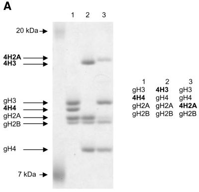
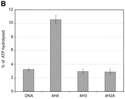
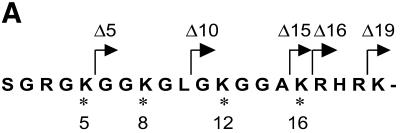
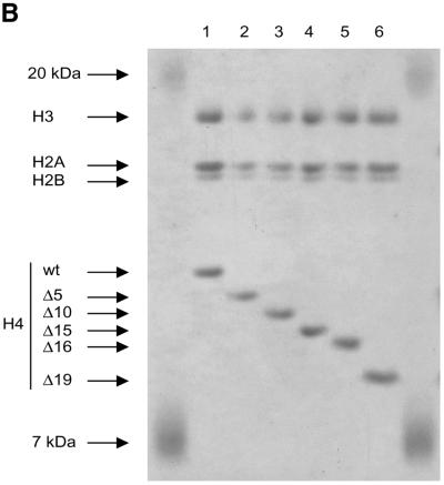
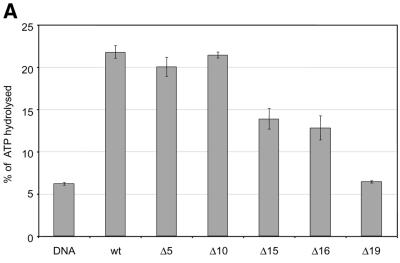
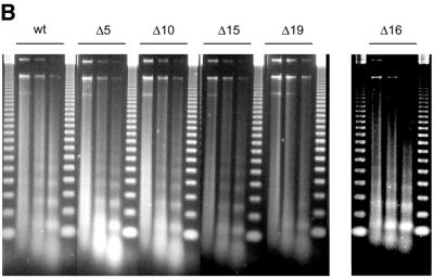
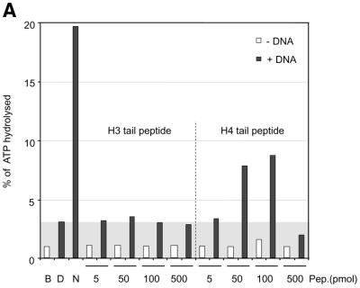
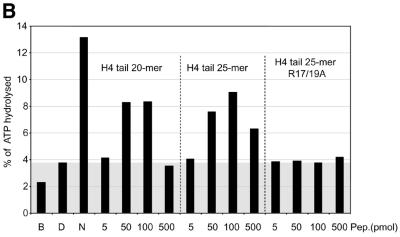
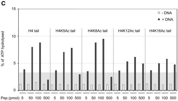
The ATPase ISWI is the catalytic core of several nucleosome remodeling complexes, which are able to alter histone-DNA interactions within nucleosomes such that the sliding of histone octamers on DNA is facilitated. Dynamic nucleosome repositioning may be involved in the assembly of chromatin with regularly spaced nucleosomes and accessible regulatory sequence elements. The mechanism that underlies nucleosome sliding is largely unresolved. We recently discovered that the N-terminal 'tail' of histone H4 is critical for nucleosome remodeling by ISWI. If deleted, nucleosomes are no longer recognized as substrates and do not stimulate the ATPase activity of ISWI. We show here that the H4 tail is part of a more complex recognition epitope which is destroyed by grafting the H4 N-terminus onto other histones. We mapped the H4 tail requirement to a hydrophilic patch consisting of the amino acids R17H18R19 localized at the base of the tail. These residues have been shown earlier to contact nucleosomal DNA, suggesting that ISWI recognizes an 'epitope' consisting of the DNA-bound H4 tail. Consistent with this hypothesis, the ISWI ATPase is stimulated by isolated H4 tail peptides ISWI only in the presence of DNA. Acetylation of the adjacent K12 and K16 residues impairs substrate recognition by ISWI.


Figures

Figure 1
Figure 1
Grafting the histone H4 N-terminus on H3 and H2A prevents substrate recognition by ISWI. (A) Analysis of chimeric histone octamers. Histone octamers were refolded from sets of variant histones as indicated on the right. The globular portions of the histones (39) are indicated with the prefix ‘g’. gH3, histone H3 from K27 to A135; gH4, histone H4 from K20 to G102; gH2A, histone H2A from K13 to K118; and gH2B, histone H2B from K27 to K125. Those histones to which the H4 N-terminus was added (see Materials and Methods) are indicated with the prefix 4. Histones were analyzed by SDS–PAGE in an 18% gel and stained with Coomassie Blue. The migration of the histones and markers are indicated on the left. (B) ATPase assays with variant histone octamers. The histones shown in (A) were reconstituted into nucleosomes by NAP-1 assisted assembly in the presence of ISWI. A control reaction contained only DNA in the absence of histones. The ATPase activity of ISWI during the reaction is displayed as the percentage of ATP hydrolyzed during the assay. The bars represent the average of three independent experiments, and the variability is indicated by the error bars.
Figure 1
Figure 1
Grafting the histone H4 N-terminus on H3 and H2A prevents substrate recognition by ISWI. (A) Analysis of chimeric histone octamers. Histone octamers were refolded from sets of variant histones as indicated on the right. The globular portions of the histones (39) are indicated with the prefix ‘g’. gH3, histone H3 from K27 to A135; gH4, histone H4 from K20 to G102; gH2A, histone H2A from K13 to K118; and gH2B, histone H2B from K27 to K125. Those histones to which the H4 N-terminus was added (see Materials and Methods) are indicated with the prefix 4. Histones were analyzed by SDS–PAGE in an 18% gel and stained with Coomassie Blue. The migration of the histones and markers are indicated on the left. (B) ATPase assays with variant histone octamers. The histones shown in (A) were reconstituted into nucleosomes by NAP-1 assisted assembly in the presence of ISWI. A control reaction contained only DNA in the absence of histones. The ATPase activity of ISWI during the reaction is displayed as the percentage of ATP hydrolyzed during the assay. The bars represent the average of three independent experiments, and the variability is indicated by the error bars.
Figure 2
Figure 2
Characterization of histone octamers harboring systematic deletions of the H4 N-terminus. (A) Amino acid sequence of the histone H4 N-terminal ‘tail’ domain. The endpoints of the deletions are indicated. Asterisks indicate the lysine residues that can be acetylated in vivo. (B) Histone octamers were refolded from histones H3, H2A, H2B and a series of H4 variants from which N-terminal amino acids were sequentially deleted. The histones were analyzed as in Figure 1. The identity of the bands is indicated on the left.
Figure 2
Figure 2
Characterization of histone octamers harboring systematic deletions of the H4 N-terminus. (A) Amino acid sequence of the histone H4 N-terminal ‘tail’ domain. The endpoints of the deletions are indicated. Asterisks indicate the lysine residues that can be acetylated in vivo. (B) Histone octamers were refolded from histones H3, H2A, H2B and a series of H4 variants from which N-terminal amino acids were sequentially deleted. The histones were analyzed as in Figure 1. The identity of the bands is indicated on the left.
Figure 3
Figure 3
Identification of crucial determinants for ISWI function in the H4 N-terminus. (A) ATPase activity of ISWI in the presence of the variant nucleosomes shown in Figure 2. ATPase activity is expressed as the percentage of ATP hydrolyzed during the assay. The bars represent the average of three independent experiments, the variability is indicated by the error bars. (B) Nucleosome ‘spacing’ activity of ISWI in the presence of the variant nucleosomes shown in Figure 2. Removal of the amino acids R17H18R19 abolishes the ‘spacing’ capacity of ISWI. The histone octamers were reconstituted into nucleosomes on plasmid DNA by NAP-1 assisted assembly in the presence of ISWI and subjected to MNase digestion. Chromatinized DNA (300 ng) was digested with 0.2 U of MNase for 20, 50 and 110 s. The resulting DNA fragments were purified and visualized by agarose gel electrophoresis and SybrGold“ staining. The marker is a 123 bp DNA ladder.
Figure 3
Figure 3
Identification of crucial determinants for ISWI function in the H4 N-terminus. (A) ATPase activity of ISWI in the presence of the variant nucleosomes shown in Figure 2. ATPase activity is expressed as the percentage of ATP hydrolyzed during the assay. The bars represent the average of three independent experiments, the variability is indicated by the error bars. (B) Nucleosome ‘spacing’ activity of ISWI in the presence of the variant nucleosomes shown in Figure 2. Removal of the amino acids R17H18R19 abolishes the ‘spacing’ capacity of ISWI. The histone octamers were reconstituted into nucleosomes on plasmid DNA by NAP-1 assisted assembly in the presence of ISWI and subjected to MNase digestion. Chromatinized DNA (300 ng) was digested with 0.2 U of MNase for 20, 50 and 110 s. The resulting DNA fragments were purified and visualized by agarose gel electrophoresis and SybrGold“ staining. The marker is a 123 bp DNA ladder.
Figure 4
Figure 4
Interaction of H4 tail peptides with DNA reconstitutes a minimal determinant for ISWI response. (A) ISWI ATPase assays in the presence of various substrates. B, buffer; D, DNA; N, nucleosomes; H3 or H4 tail peptides in the indicated amounts (5–500 pmol) in the absence (–) or presence (+) of DNA. The ATPase activity is expressed as the percentage of ATP hydrolyzed during the assay. The ATPase activity level elicited by free DNA is extended through the figure as a gray background, for reference. This result has been obtained consistently in a number of independent experiments under different conditions. (B) R17/R19 within the histone H4 tail are crucial for ISWI ATPase activity. A 25mer H4 tail peptide TGRGKGGKGLGKGGAKRHRKVLRDC induces the ATPase of ISWI as well as the 20mer used in (A). A variant 25mer H4 tail peptide in which R17 and R19 were substituted by alanines does not trigger ATPase acivity. All reactions contained DNA. (C) ISWI ATPase activity is sensitive to site-specific acetylation of histone H4 tail. The data are presented as in (A).
Figure 4
Figure 4
Interaction of H4 tail peptides with DNA reconstitutes a minimal determinant for ISWI response. (A) ISWI ATPase assays in the presence of various substrates. B, buffer; D, DNA; N, nucleosomes; H3 or H4 tail peptides in the indicated amounts (5–500 pmol) in the absence (–) or presence (+) of DNA. The ATPase activity is expressed as the percentage of ATP hydrolyzed during the assay. The ATPase activity level elicited by free DNA is extended through the figure as a gray background, for reference. This result has been obtained consistently in a number of independent experiments under different conditions. (B) R17/R19 within the histone H4 tail are crucial for ISWI ATPase activity. A 25mer H4 tail peptide TGRGKGGKGLGKGGAKRHRKVLRDC induces the ATPase of ISWI as well as the 20mer used in (A). A variant 25mer H4 tail peptide in which R17 and R19 were substituted by alanines does not trigger ATPase acivity. All reactions contained DNA. (C) ISWI ATPase activity is sensitive to site-specific acetylation of histone H4 tail. The data are presented as in (A).

References

    1. Kingston R.E. and Narlikar,G.J. (1999) ATP-dependent remodeling and acetylation as regulators of chromatin fluidity. Genes Dev., 13, 2339–2352. - PubMed
    1. Peterson C.L. (2000) ATP-dependent chromatin remodeling: going mobile. FEBS Lett., 476, 68–72. - PubMed
    1. Havas K., Whitehouse,I. and Owen-Hughes,T. (2001) ATP-dependent chromatin remodeling activities. Cell. Mol. Life Sci., 58, 673–682. - PMC - PubMed
    1. Vignali M., Hassan,A.H., Neely,K.E. and Workman,J.L. (2000) ATP-dependent chromatin-remodeling complexes. Mol. Cell. Biol., 20, 1899–1910. - PMC - PubMed
    1. Becker P.B. and Hörz,W. (2002) ATP-dependent nucleosome remodeling. Annu. Rev. Biochem., in press. - PubMed

Publication types

MeSH terms