. 2002 Feb 1;30(3):685-94.
doi: 10.1093/nar/30.3.685.

Identification, cloning and characterization of a new DNA-binding protein from the hyperthermophilic methanogen Methanopyrus kandleri

Affiliations

Identification, cloning and characterization of a new DNA-binding protein from the hyperthermophilic methanogen Methanopyrus kandleri

Nikolai A Pavlov et al. Nucleic Acids Res. .

Abstract

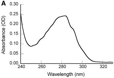
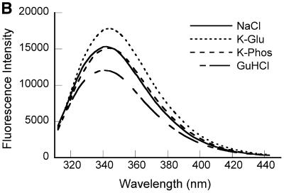
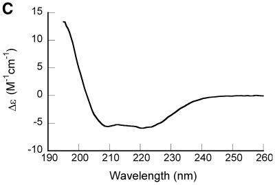
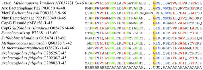
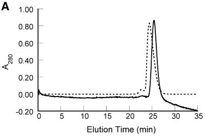
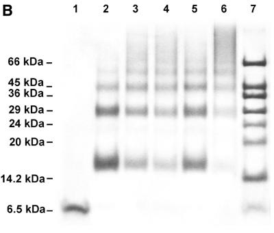
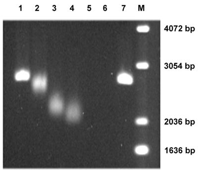
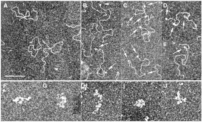
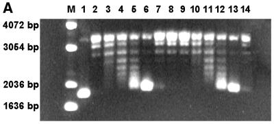
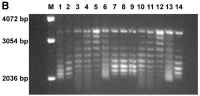
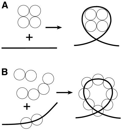
Three novel DNA-binding proteins with apparent molecular masses of 7, 10 and 30 kDa have been isolated from the hyperthermophilic methanogen Methanopyrus kandleri. The proteins were identified using a blot overlay assay that was modified to emulate the high ionic strength intracellular environment of M.kandleri proteins. A 7 kDa protein, named 7kMk, was cloned and expressed in Escherichia coli. As indicated by CD spectroscopy and computer-assisted structure prediction methods, 7kMk is a substantially alpha-helical protein possibly containing a short N-terminal beta-strand. According to analytical gel filtration chromatography and chemical crosslinking, 7kMk exists as a stable dimer, susceptible to further oligomerization. Electron microscopy showed that 7kMk bends DNA and also leads to the formation of loop-like structures of approximately 43.5 +/- 3.5 nm (136 +/- 11 bp for B-form DNA) circumference. A topoisomerase relaxation assay demonstrated that looped DNA is negatively supercoiled under physiologically relevant conditions (high salt and temperature). A BLAST search did not yield 7kMk homologs at the amino acid sequence level, but based on a multiple alignment with ribbon-helix-helix (RHH) transcriptional regulators, fold features and self-association properties of 7kMk we hypothesize that it could be related to RHH proteins.


Figures

Figure 1
Figure 1
Spectroscopic characterization of purified recombinant 7kMk protein. (A) UV absorption spectrum of 7kMk protein (0.24 mg/ml) in NB buffer. (B) Fluorescence emission spectra of 7kMk (0.14 mg/ml) acquired at an excitation wavelength of 295 nm in buffers NB (‘NaCl’), 1 M K2HPO4/KH2PO4 pH 7.2 (‘K-Phos’), GB (‘K-Glu’) and NB supplemented with 6 M guanidine HCl (‘GuHCl’). (C) CD spectrum of 0.78 mg/ml 7kMk in a buffer containing 1 M NaCl, 10 mM NaH2PO4/Na2HPO4, pH 7.2, obtained with a 0.1 mm path length cuvette.
Figure 1
Figure 1
Spectroscopic characterization of purified recombinant 7kMk protein. (A) UV absorption spectrum of 7kMk protein (0.24 mg/ml) in NB buffer. (B) Fluorescence emission spectra of 7kMk (0.14 mg/ml) acquired at an excitation wavelength of 295 nm in buffers NB (‘NaCl’), 1 M K2HPO4/KH2PO4 pH 7.2 (‘K-Phos’), GB (‘K-Glu’) and NB supplemented with 6 M guanidine HCl (‘GuHCl’). (C) CD spectrum of 0.78 mg/ml 7kMk in a buffer containing 1 M NaCl, 10 mM NaH2PO4/Na2HPO4, pH 7.2, obtained with a 0.1 mm path length cuvette.
Figure 2
Figure 2
Multiple alignment of 7kMk, RHH proteins with solved crystal structures (names in bold) and several putative archaeal RHH proteins. Hydrophobic (LIYFWVMA), polar (STNREQHD) and small (GASNSTCP) residues conserved in >80% of the sequences are colored red, green and blue, respectively. The conserved glycine residue of the turn connecting α-helices is colored violet and highlighted in yellow. The names of proteins, origins, accession codes and numbering of residues are indicated. Elements of secondary structure are given below the alignment. A, α-helix; B, β-strand; U, unordered.
Figure 3
Figure 3
Self-association behavior of 7kMk protein. (A) Elution profiles of gel filtration chromatography. 7kMk was loaded onto a Superdex 75 column at concentrations of 5.4 µM (continuous line) and 540 µM (broken line). (B) Chemical crosslinking of 7kMk protein. 7kMk samples diluted with NB buffer to final concentrations of 5 µM (lane 2), 20 µM (lanes 3–5) and 100 µM (lane 6) were digested with glutaraldehyde at final concentrations of 0.03% (lane 5), 0.1% (lanes 2, 4 and 6) and 0.3% (lane 3). Lane 1, 7kMk without fixation.
Figure 3
Figure 3
Self-association behavior of 7kMk protein. (A) Elution profiles of gel filtration chromatography. 7kMk was loaded onto a Superdex 75 column at concentrations of 5.4 µM (continuous line) and 540 µM (broken line). (B) Chemical crosslinking of 7kMk protein. 7kMk samples diluted with NB buffer to final concentrations of 5 µM (lane 2), 20 µM (lanes 3–5) and 100 µM (lane 6) were digested with glutaraldehyde at final concentrations of 0.03% (lane 5), 0.1% (lanes 2, 4 and 6) and 0.3% (lane 3). Lane 1, 7kMk without fixation.
Figure 4
Figure 4
EMSA of 7kMk–DNA complexes. BamHI linearized pUC19 DNA (150 ng) was incubated with various concentrations of 7kMk in GB buffer at 70°C for 30 min. The Rw (protein:DNA weight ratio) values of the samples loaded in lanes 1–7 were 0.5, 1, 1.5, 2, 3, 10 and 0, respectively. M, 1 kb DNA ladder (Gibco BRL). Electrophoresis was performed in 1.5% agarose at 1.5 V/cm for 16 h in TBE buffer containing 100 mM Na-Glu.
Figure 5
Figure 5
EM of the complexes formed of 7kMk with pUC19/BamHI DNA. Complexes were obtained after incubation in the presence of 1 M K-Glu at 70°C at a Rw of 0 (A), 1.5 (BE) and 10 (FJ). Arrows indicate DNA loops and bends. The scale bar represents 200 nm.
Figure 6
Figure 6
DNA topology assay of 7kMk protein. Relaxed pUC19 (300 ng) was incubated with various concentrations of 7kMk in GB buffer at 70°C for 30 min. 7kMk was added at a Rw of 0 (lane 10), 0.4 (lane 3), 0.8 (lanes 4 and 11), 1.6 (lanes 5 and 12), 3.3 (lanes 6 and 13) 5 (lane 7), 6.7 (lanes 8 and 14) and 12 (lane 9). The resulting complexes were digested with topoisomerase V (100 ng) at the same temperature for 15 min (lanes 3–10) or 1 h (lanes 11–14). Lanes M, 1 and 2 were a 1 kb DNA ladder (Gibco BRL), negatively supercoiled pUC19 DNA isolated from E.coli and relaxed pUC19 DNA, respectively. The reaction products were analyzed by 1.5% agarose gel electrophoresis in TBE buffer (A) or TBE buffer containing 1 µg/ml chloroquine (B) at 1.5 V/cm for 16 h, stained with ethidium bromide and digitized.
Figure 6
Figure 6
DNA topology assay of 7kMk protein. Relaxed pUC19 (300 ng) was incubated with various concentrations of 7kMk in GB buffer at 70°C for 30 min. 7kMk was added at a Rw of 0 (lane 10), 0.4 (lane 3), 0.8 (lanes 4 and 11), 1.6 (lanes 5 and 12), 3.3 (lanes 6 and 13) 5 (lane 7), 6.7 (lanes 8 and 14) and 12 (lane 9). The resulting complexes were digested with topoisomerase V (100 ng) at the same temperature for 15 min (lanes 3–10) or 1 h (lanes 11–14). Lanes M, 1 and 2 were a 1 kb DNA ladder (Gibco BRL), negatively supercoiled pUC19 DNA isolated from E.coli and relaxed pUC19 DNA, respectively. The reaction products were analyzed by 1.5% agarose gel electrophoresis in TBE buffer (A) or TBE buffer containing 1 µg/ml chloroquine (B) at 1.5 V/cm for 16 h, stained with ethidium bromide and digitized.
Figure 7
Figure 7
Two models of 7kMk–DNA complex formation. (A) Nucleosome-like wrapping of DNA by the 7kMk oligomer. (B) Cooperative binding of 7kMk to the DNA molecule resulting in DNA bending and looping.

References

    1. Sandman K., Krzycki,J.A., Dobrinski,B., Lurz,R. and Reeve,J.N. (1990) HMf, a DNA-binding protein isolated from the hyperthermophilic archaeon Methanothermus fervidus, is most closely related to histones. Proc. Natl Acad. Sci. USA, 87, 5788–5791. - PMC - PubMed
    1. Sandman K. and Reeve,J.N. (2000) Structure and functional relationships of archaeal and eukaryal histones and nucleosomes. Arch. Microbiol., 173, 165–169. - PubMed
    1. Langer D., Hain,J., Thuriaux,P. and Zillig,W. (1995) Transcription in archaea: similarity to that in eucarya. Proc. Natl Acad. Sci. USA, 92, 5768–5772. - PMC - PubMed
    1. Rowlands T., Baumann,P. and Jackson,S.P. (1994) The TATA-binding protein: a general transcription factor in eukaryotes and archaebacteria. Science, 264, 1326–1329. - PubMed
    1. Ouzounis C. and Sander,C. (1992) TFIIB, an evolutionary link between the transcription machineries of archaebacteria and eukaryotes. Cell, 71, 189–190. - PubMed

Publication types

MeSH terms

Associated data