Comparative Study
. 2002 Feb;47(2):372-83.
doi: 10.1002/mrm.10051.

Phase-sensitive inversion recovery for detecting myocardial infarction using gadolinium-delayed hyperenhancement

Affiliations
Comparative Study

Phase-sensitive inversion recovery for detecting myocardial infarction using gadolinium-delayed hyperenhancement

Peter Kellman et al. Magn Reson Med. 2002 Feb.

Abstract

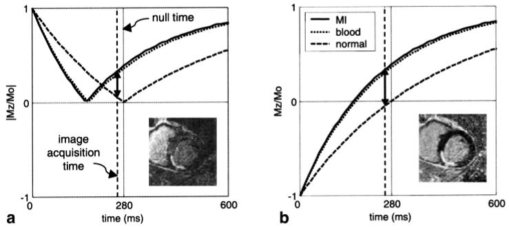
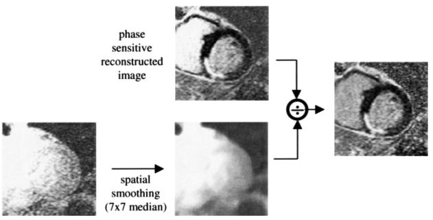
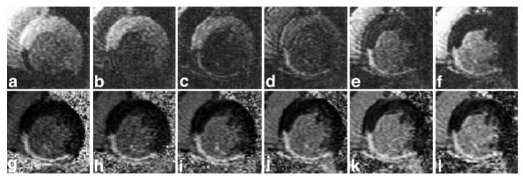
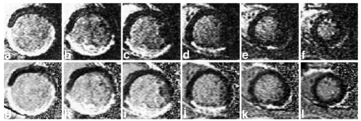
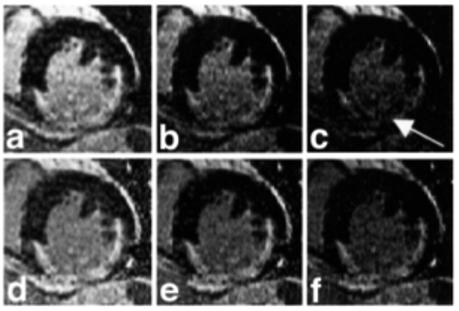
After administration of gadolinium, infarcted myocardium exhibits delayed hyperenhancement and can be imaged using an inversion recovery (IR) sequence. The performance of such a method when using magnitude-reconstructed images is highly sensitive to the inversion recovery time (TI) selected. Using phase-sensitive reconstruction, it is possible to use a nominal value of TI, eliminate several breath-holds otherwise needed to find the precise null time for normal myocardium, and achieve a consistent contrast. Phase-sensitive detection is used to remove the background phase while preserving the sign of the desired magnetization during IR. Experimental results are presented which demonstrate the benefits of both phase-sensitive IR image reconstruction and surface coil intensity normalization for detecting myocardial infarction (MI). The phase-sensitive reconstruction method reduces the variation in apparent infarct size that is observed in the magnitude images as TI is changed. Phase-sensitive detection also has the advantage of decreasing the sensitivity to changes in tissue T(1) with increasing delay from contrast agent injection.


Figures

FIG. 1
FIG. 1
Plots of signal intensity vs. TI for (a) magnitude and (b) phase-sensitive detection for MI (solid), blood (dotted), and normal myocardium (dashed), using nominal values of T1 at 15 min following a double dose of contrast agent. Example images correspond to acquiring images earlier than the null time for normal myocardium. The solid lines with double arrows depict the contrast between the MI and the normal myocardium.
FIG. 2
FIG. 2
Pulse sequence diagram for gated, segmented k-space acquisition of IR and reference images using low flip-angle readouts. Data for IR and reference images are collected alternately every other heartbeat.
FIG. 3
FIG. 3
Block diagram showing the phased-array phase-sensitive reconstruction of IR image using a separate reference image acquired after magnetization recovery.
FIG. 4
FIG. 4
Method for adaptively estimating phased-array combiner coefficients using the multicoil complex reference images.
FIG. 5
FIG. 5
Surface coil intensity normalization.
FIG. 6
FIG. 6
Probability distributions of signal-plus-noise for (a) magnitude and (b) phase-sensitive detection for phased array with four coils.
FIG. 7
FIG. 7
Short-axis images at varied TIs for a patient with inferior MI. Magnitude (top row) and normalized phase-sensitive (bottom row) detection for TI = 175, 200, 225, 250, 275, and 300 ms from left to right. The appearance and contrast are variable for the magnitude-reconstructed images, while they are consistent for the normalized phase-sensitive reconstruction.
FIG. 8
FIG. 8
Short-axis stack images for another patient with inferior MI, comparing magnitude (top row) and normalized phase-sensitive (bottom row) detection for six slices, acquired over approximately 4 min for columns from left to right (basal to apical). A slight degradation in contrast is seen in the magnitude images for the (a and b) basal slices, for which the TI is slightly less than the null time for the normal myocardium, while a uniform contrast is achieved in the (g-l) phase-sensitive images acquired simultaneously.
FIG. 9
FIG. 9
Example short-axis images shown at different display signal intensity levels (window and level) illustrate the uniformity across the MI region (a-c) before and (d-f) after surface coil intensity correction.
FIG. 10
FIG. 10
Output SNR for magnitude and normalized phase-sensitive images for (a) measured phantom data SNR vs. |Mz/Mo| for three values of reference image SNR, and (b) Monte Carlo simulation of output SNR vs. input SNR for approximately the same three values of reference SNR plus ideal noise-free reference. The bold lines correspond to magnitude detection. The reference SNR for phase detection is 4, 8, and 16, for diamond, circle, and squares, respectively. The simulated ideal noiseless reference case shown in b is plotted using triangles. The gray shaded region in b corresponds to typical SNR values for LV blood pool or MI, for which the reference SNR is greater than or equal to the SNR of the IR image.
FIG. 11
FIG. 11
CNR between LV blood pool and normal myocardium for both magnitude and phase-sensitive reconstruction methods measured for 20 patients.
FIG. 12
FIG. 12
Signal intensity of phantom IR image vs. TI for several values of reference image RF readout flip angle, validating minimal loss in the IR image due to reference image acquisition.

References

    1. Kim RJ, Fieno DS, Parrish TB, Harris K, Chen EL, Simonetti O, Bundy J, Finn JP, Klocke FJ, Judd RM. Relationship of MRI delayed contrast enhancement to irreversible injury, infarct age, and contractile function. Circulation. 1999;100:1992–2002. - PubMed
    1. Kim RJ, Wu E, Rafael A, Chen EL, Parker MA, Simonetti O, Klocke FJ, Bonow RO, Judd RM. The use of contrast-enhanced magnetic resonance imaging to identify reversible myocardial dysfunction. N Engl J Med. 2000;343:1445–1453. - PubMed
    1. Kim RJ, Hillenbrand HB, Judd RM. Evaluation of myocardial viability by MRI. Herz. 2000;25:417–430. - PubMed
    1. Wu KC, Rochitte CE, Lima JA. Magnetic resonance imaging in acute myocardial infarction. Curr Opin Cardiol. 1999;14:480–484. - PubMed
    1. Simonetti OP, Kim RJ, Fieno DS, Hillenbrand HB, Wu E, Bundy JM, Finn JP, Judd RM. An improved MR imaging technique for the visualization of myocardial infarction. Radiology. 2001;218:215–223. - PubMed

Publication types