

Changes in markers of bone metabolism during dexamethasone treatment for chronic lung disease in preterm infants
- PMID: 11815549
- PMCID: PMC1721351
- DOI: 10.1136/fn.86.1.f49
Changes in markers of bone metabolism during dexamethasone treatment for chronic lung disease in preterm infants
Abstract
Aim: To characterise the change in serum and urinary bone markers in the early postnatal period, and to assess the effect of systemic corticosteroid on bone metabolism in preterm infants.
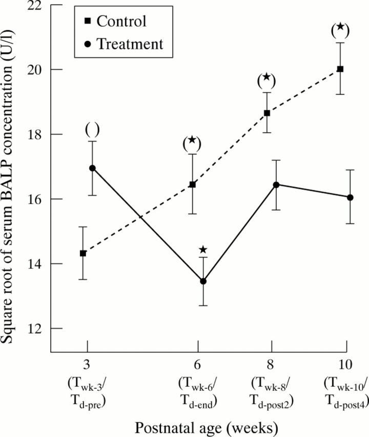
Methods: Bone formation was quantified by measurement of serum concentrations of bone specific alkaline phosphatase (BALP) and osteocalcin. Bone resorption was measured by monitoring creatinine adjusted urinary deoxypyridinoline (Dpd) concentration. Blood and urinary samples were collected from corticosteroid treated infants (n = 19) immediately before the start (T(d-pre)), three weeks after the start (T(d-end)), and two (T(d-post2)) and four weeks (T(d-post4)) after the end of the dexamethasone course. Untreated patients (n = 30) had specimens taken at week 3 (T(wk-3)), 6 (T(wk-6)), 8 (T(wk-8)), and 10 (T(wk-10)) of postnatal age.
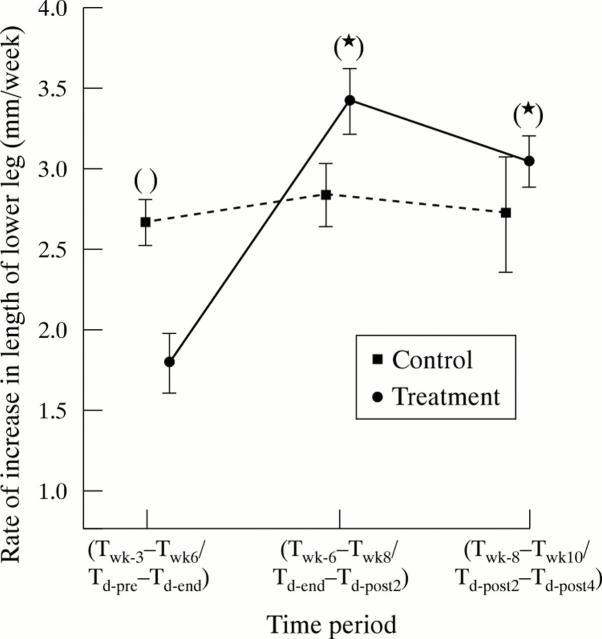
Results: Serum concentrations of BALP and osteocalcin at T(d-end) were significantly lower than pretreatment levels and the levels at the corresponding time point (T(wk-6)) of the non-treatment group. In contrast, urinary Dpd concentration at T(d-end) was not significantly decreased compared with the pretreatment level. However, it was significantly lower than the urinary Dpd concentration at T(wk-6) of the non-treatment group. The rate of increase in lower leg length was significantly higher in the non-treatment group between weeks 3 and 6 than in the corresponding period during dexamethasone treatment in the corticosteroid group.
Conclusion: Systemic corticosteroid causes appreciable suppression of serum BALP and osteocalcin and, to a lesser extent, urinary Dpd. The results suggest that corticosteroid inhibits bone growth mainly by decreasing bone formation.
Figures


Similar articles
-
Short-term effects on linear growth and bone turnover in children randomized to receive prednisolone or dexamethasone.Clin Endocrinol (Oxf). 2002 Aug;57(2):185-91. doi: 10.1046/j.1365-2265.2002.01580.x. Clin Endocrinol (Oxf). 2002. PMID: 12153596 Clinical Trial.
-
Changes in serum leptin concentration after corticosteroid treatment in preterm infants.Acta Paediatr. 2002;91(6):684-90. doi: 10.1080/080352502760069124. Acta Paediatr. 2002. PMID: 12162603 Clinical Trial.
-
Changes in (markers of) bone metabolism during high dose corticosteroid pulse treatment in patients with rheumatoid arthritis.Ann Rheum Dis. 1996 May;55(5):288-93. doi: 10.1136/ard.55.5.288. Ann Rheum Dis. 1996. PMID: 8660101 Free PMC article.
-
Effects of dexamethasone treatment on bone and collagen turnover in preterm infants with chronic lung disease.Pediatr Res. 2000 Aug;48(2):155-62. doi: 10.1203/00006450-200008000-00007. Pediatr Res. 2000. PMID: 10926289 Clinical Trial.
-
Effect of different doses of vitamin D on osteocalcin and deoxypyridinoline in preterm infants.Pediatr Int. 2008 Apr;50(2):204-7. doi: 10.1111/j.1442-200X.2008.02553.x. Pediatr Int. 2008. PMID: 18353060 Clinical Trial.
Cited by
-
Inhibition of ERK promotes collagen gel compaction and fibrillogenesis to amplify the osteogenesis of human mesenchymal stem cells in three-dimensional collagen I culture.Stem Cells Dev. 2009 Mar;18(2):331-41. doi: 10.1089/scd.2008.0075. Stem Cells Dev. 2009. PMID: 18491946 Free PMC article.
-
Vitamin D receptor polymorphisms and growth until adulthood after very premature birth.J Bone Miner Metab. 2016 Sep;34(5):564-70. doi: 10.1007/s00774-015-0697-8. Epub 2015 Jul 28. J Bone Miner Metab. 2016. PMID: 26212485
-
Dissection of the osteogenic effects of laminin-332 utilizing specific LG domains: LG3 induces osteogenic differentiation, but not mineralization.Exp Cell Res. 2008 Feb 15;314(4):763-73. doi: 10.1016/j.yexcr.2007.12.007. Epub 2008 Jan 22. Exp Cell Res. 2008. PMID: 18206871 Free PMC article.
-
The Clinical and Biochemical Predictors of Bone Mass in Preterm Infants.PLoS One. 2016 Nov 2;11(11):e0165727. doi: 10.1371/journal.pone.0165727. eCollection 2016. PLoS One. 2016. PMID: 27806112 Free PMC article.
-
Osteogenic differentiation of mesenchymal stem cells in defined protein beads.J Biomed Mater Res B Appl Biomater. 2008 Oct;87(1):213-21. doi: 10.1002/jbm.b.31098. J Biomed Mater Res B Appl Biomater. 2008. PMID: 18431753 Free PMC article.
References
MeSH terms
Substances
LinkOut - more resources
Full Text Sources
Medical