. 2002 Feb 4;156(3):419-24.
doi: 10.1083/jcb.200111002. Epub 2002 Jan 28.

Condensin and cohesin display different arm conformations with characteristic hinge angles

Affiliations

Condensin and cohesin display different arm conformations with characteristic hinge angles

David E Anderson et al. J Cell Biol. .

Abstract

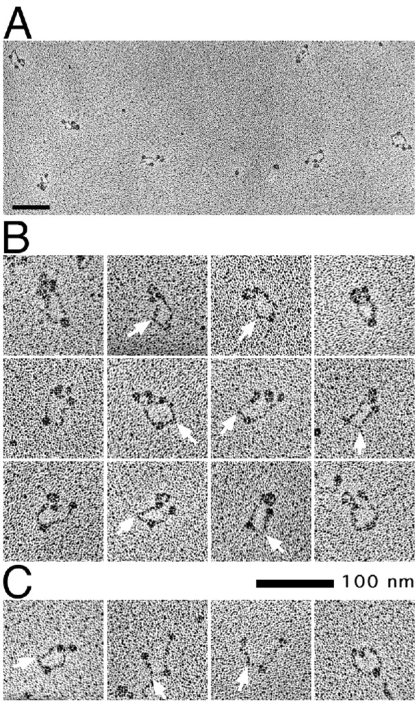
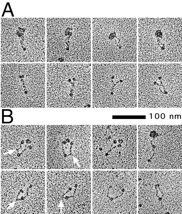
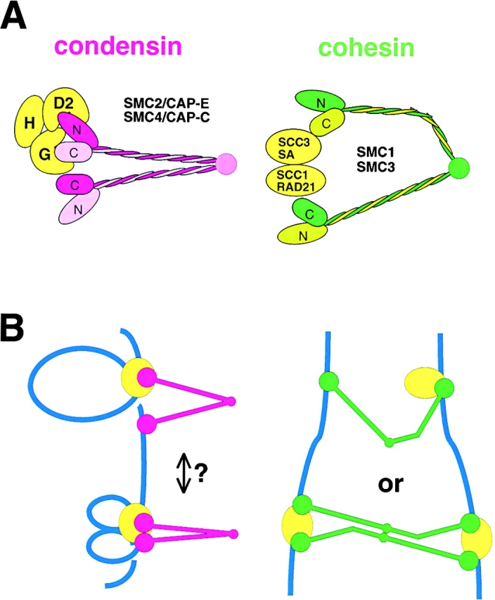
Structural maintenance of chromosomes (SMC) proteins play central roles in higher-order chromosome dynamics from bacteria to humans. In eukaryotes, two different SMC protein complexes, condensin and cohesin, regulate chromosome condensation and sister chromatid cohesion, respectively. Each of the complexes consists of a heterodimeric pair of SMC subunits and two or three non-SMC subunits. Previous studies have shown that a bacterial SMC homodimer has a symmetrical structure in which two long coiled-coil arms are connected by a flexible hinge. A catalytic domain with DNA- and ATP-binding activities is located at the distal end of each arm. We report here the visualization of vertebrate condensin and cohesin by electron microscopy. Both complexes display the two-armed structure characteristic of SMC proteins, but their conformations are remarkably different. The hinge of condensin is closed and the coiled-coil arms are placed close together. In contrast, the hinge of cohesin is wide open and the coiled-coils are spread apart from each other. The non-SMC subunits of both condensin and cohesin form a globular complex bound to the catalytic domains of the SMC heterodimers. We propose that the "closed" conformation of condensin and the "open" conformation of cohesin are important structural properties that contribute to their specialized biochemical and physiological functions.


Figures

Figure 1.
Figure 1.
Electron micrographs of the human condensin complexes. (A) An example field of molecules. (B) The structure of holocomplexes can be classified into three major groups on the basis of the configuration of their coiled-coil arms: ‘folded-rod’ (first row) and ‘ends-split’ (second row)—a subset of which have a bend in the coils (third row)—and ‘coils-spread’ (fourth row). The last panel in the fourth row is an example of a rare ‘open-V’. (C) The two major forms of SMC heterodimers (hSMC2-hSMC4) are again folded-rod (first row) and ends-split (second row), some with bent coils (third row). Open-V configurations are rare (last panel, second row). The hinge domain is indicated by an arrow on the open-V molecules. B and C are the same magnification. Bars, 100 nm.
Figure 2.
Figure 2.
Electron micrographs of the human cohesin complexes. (A) An example field of molecules. (B) The structure of holocomplexes can be classified into two major groups on the basis of the configuration of their coiled-coil arms. In the first row the molecules are in a ‘coils-spread’ conformation, with their catalytic domains and the non-SMC proteins mostly superimposed. In the second and third rows the molecules are in an open-V conformation with the heads somewhat separated. The non-SMC complex appears either as a separate globule between the catalytic domains (second row), or bound to one of them (third row). A kink is often observed at a fixed position in one of the two coiled-coil arms (indicated by arrows). (C) SMC heterodimers (hSMC1-hSMC3) show mostly open-V configurations. Again, one of the coiled-coil arms often displays a kink (indicated by arrows). B and C are the same magnification. Bars, 100 nm.
Figure 3.
Figure 3.
Electron micrographs of the Xenopus condensin and cohesin complexes. (A) Holocomplexes of Xenopus condensin (first row) and heterodimers of XCAP-E/XSMC2 and XCAP-C/XSMC4 (second row). (B) Holocomplexes of Xenopus cohesin (first row) and heterodimers of XSMC1 and XSMC3 (second row). Arrows in B indicate the position of the kink in the coiled-coil arm. Bar, 100 nm.
Figure 4.
Figure 4.
Models. (A) Molecular architecture of condensin and cohesin. N and C indicate NH2-terminal and COOH-terminal nonhelical domains, respectively, of the SMC subunits. By analogy to BsSMC, we assume here that the coiled-coil arms of the eukaryotic SMC heterodimers are arranged into an antiparallel fashion. The relative positions of the non-SMC subunits (shown in yellow) are arbitrary. (B) Hypothetical models of the interactions of condensin (left) and cohesin (right) with DNA.

References

    1. Anderson, D.E., K.M. Trujillo, P. Sung, and H.P. Erickson. 2001. Structure of the Rad50/Mre11 DNA repair complex from Saccharomyces cerevisiae by electron microscopy. J. Biol. Chem. 276:37027–37033. - PubMed
    1. Birkenbihl, R.P., and S. Subramani. 1992. Cloning and characterization of rad21, an essential gene of Schizosaccharomyces pombe involved in DNA double-strand-break repair. Nucleic Acids Res. 20:6605–6611. - PMC - PubMed
    1. Cobbe, N., and M.M. Heck. 2000. SMCs in the world of chromosome biology: from prokaryotes to higher eukaryotes. J. Struct. Biol. 129:123–143. - PubMed
    1. Dej, K.J., and T.L. Orr-Weaver. 2000. Separation anxiety at the centromere. Trends Cell Biol. 10:392–399. - PubMed
    1. Fowler, W.E., and H.P. Erickson. 1979. Trinodular structure of fibrinogen. Confirmation by both shadowing and negative stain electron microscopy. J. Mol. Biol. 134:241–249. - PubMed

Publication types

MeSH terms