. 2002 Feb 5;99(3):1621-6.
doi: 10.1073/pnas.022646599. Epub 2002 Jan 29.

Cultured rat hippocampal neural progenitors generate spontaneously active neural networks

Affiliations

Cultured rat hippocampal neural progenitors generate spontaneously active neural networks

Sanjay K Mistry et al. Proc Natl Acad Sci U S A. .

Abstract

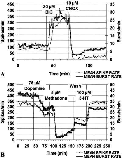
We previously demonstrated that the neural cell adhesion molecule (N-CAM) inhibited the proliferation of cultured rat hippocampal progenitor cells and increased the number of neurons generated. We demonstrate here that the continued presence of fibroblast growth factor 2 along with N-CAM or brain-derived neurotrophic factor over 12 days of culture greatly increased the number of both progenitors and neurons. These progenitor-derived neurons expressed neurotransmitters, neurotransmitter receptors, and synaptic proteins in vitro consistent with those expressed in the mature hippocampus. Progenitor cells cultured on microelectrode plates formed elaborate neural networks that exhibited spontaneously generated action potentials after 21 days. This activity was observed only in cultures grown in the presence of fibroblast growth factor 2 and either N-CAM or brain-derived neurotrophic factor. Analysis of neuronal activity after various pharmacological treatments indicated that the networks formed functional GABAergic and glutamatergic synapses. We conclude that mitogenic growth factors can synergize with N-CAM or neurotrophins to generate spontaneously active neural networks from neural progenitors.


Figures

Figure 1
Figure 1
Effect of N-CAM and BDNF on total cell number and neuron generation. Cells were cultured as described in Materials and Methods. (A) Cell counts are the average ± SD from three experiments in duplicate. (B) The number of nestin+, MAP2B+, and GFAP+ cells at day 10 was determined by counting a minimum of 400 cells in five randomly selected fields.
Figure 2
Figure 2
Immunocytochemical staining of differentiated progenitors. Cells were cultured in the presence of N-CAM as described in Materials and Methods. After 14 days, cells were processed for immunocytochemistry for the antigens, as noted. D, 4′,6-diamidino-2-phenylindole.
Figure 3
Figure 3
Demonstration of spontaneous activity in networks of progenitor-derived neurons differentiated in the presence of N-CAM (A, B) and E18 hippocampal primary neurons (C, D) at comparable stages of in vitro development on MEAs. Vertical tick marks indicate spike times recorded from single neurons. Units of activity from separate electrodes in the MEA are displayed vertically, with A–D representing 45 s of activity in the horizontal axis.
Figure 4
Figure 4
Maturation of action potentials in progenitor-derived neuronal networks. (A) Representative figure demonstrating changes in action potential shape in a culture differentiated with BDNF (100 ng/ml) in the presence of FGF2 (at 24 d in culture). Shallow broad action potentials characteristic of initial spontaneous activity (Left) change to faster more depolarized potentials within 24 h (Right). (B) Cross-correlograms were calculated from 1,400 s of spiking activity from four single units referenced to a fifth unit. The spiking activity reveals variable latencies in firing after the reference, consistent with both mono- and polysynaptic connectivity in different units in the MEA. Horizontal dash lines represent mean firing rate calculated for a Poisson spike train.
Figure 5
Figure 5
Spontaneous activity in progenitor-derived neuronal networks is mediated through inhibitory and excitatory synapses and is modulated by receptor agonists and antagonists. (A) Disinhibition with the GABAA antagonist bicuculline (30 μM, 42 min) elicited increases in network activity within 3 min, demonstrating the presence of functional GABAergic synapses. Application of the AMPA/kainate receptor antagonist CNQX (10 μM, 75 min) produced a rapid decrease in both bursting and spiking patterns, demonstrating the presence of glutamatergic synapses. Mean spike and burst rates were calculated from the activity of 23 single units in a culture differentiated with BDNF and FGF2 (at 28 d in culture). (B) Application of dopamine (75 μM, 50 min) or methadone (5 μM, 93 min) inhibited both bursting and spiking patterns, revealing the presence of dopaminergic and opioid receptors. Application of 5-hydroxytryptamine (serotonin) (100 μM) caused an increase in burst and spike activity demonstrating the presence of serotonin receptors. Mean spike and burst rates were calculated from the activity of 33 single units in a culture differentiated with N-CAM and FGF2 (at 25 d in culture), as described above.

References

    1. Palmer T D, Takahashi J, Gage F H. Mol Cell Neurosci. 1997;8:389–404. - PubMed
    1. Shetty A K, Turner D A. J Neurobiol. 1998;35:395–425. - PubMed
    1. Vicario-Abejon C, Collin C, Tsoulfas P, McKay R D. Eur J Neurosci. 2000;12:677–688. - PubMed
    1. Sah D W, Ray J, Gage F H. J Neurobiol. 1997;32:95–110. - PubMed
    1. Tanaka T, Saito H, Matsuki N. Brain Res. 1996;723:190–195. - PubMed

Publication types

MeSH terms

Substances