

Importance of epidermal growth factor receptor signaling in establishment of adenomas and maintenance of carcinomas during intestinal tumorigenesis
- PMID: 11818567
- PMCID: PMC122223
- DOI: 10.1073/pnas.032678499
Importance of epidermal growth factor receptor signaling in establishment of adenomas and maintenance of carcinomas during intestinal tumorigenesis
Abstract
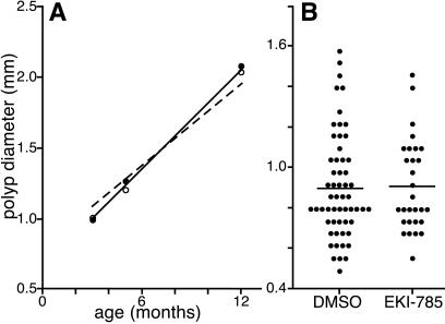
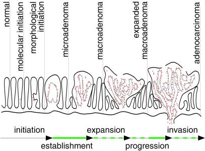
We used the hypomorphic Egfr(wa2) allele to genetically examine the impact of impaired epidermal growth factor receptor (Egfr) signaling on the Apc(Min) mouse model of familial adenomatous polyposis. Transfer of the Apc(Min) allele onto a homozygous Egfr(wa2) background results in a 90% reduction in intestinal polyp number relative to Apc(Min) mice carrying a wild-type Egfr allele. This Egfr effect is potentially synergistic with the actions of the modifier-of-min (Mom1) locus. Surprisingly, the size, expansion, and pathological progression of the polyps appear Egfr-independent. Histological examination of the ilea of younger animals revealed no differences in the number of microadenomas, the presumptive precursor lesions to gross intestinal polyps. Pharmacological inhibition with EKI-785, an Egfr tyrosine kinase inhibitor, produced similar results in the Apc(Min) model. These data suggest that normal Egfr activity is required for establishment of intestinal tumors in the Apc(Min) model between initiation and subsequent expansion of initiated tumors. The role of Egfr signaling during later stages of tumorigenesis was examined by using nude mice xenografts of two human colorectal cancer cell lines. Treatment with EKI-785 produced a dose-dependent reduction in tumor growth, suggesting that Egfr inhibitors may be useful for advanced colorectal cancer treatment.
Figures


References
-
- Gullick W J. Biochem Soc Symp. 1998;63:193–198. - PubMed
-
- Threadgill D W, Dlugosz A A, Hansen L A, Tennenbaum T, Lichti U, Yee D, LaMantia C, Mourton T, Herrup K, Harris R C, et al. Science. 1995;269:230–234. - PubMed
-
- Sibilia M, Wagner E F. Science. 1995;269:234–238. - PubMed
-
- Miettinen P J, Berger J E, Meneses J, Phung Y, Pedersen R A, Werb Z, Derynck R. Nature (London) 1995;376:337–341. - PubMed
-
- Luetteke N C, Phillips H K, Qiu T H, Copeland N G, Earp H S, Jenkins N A, Lee D C. Genes Dev. 1994;8:399–413. - PubMed
Publication types
MeSH terms
Substances
Grants and funding
LinkOut - more resources
Full Text Sources
Other Literature Sources
Molecular Biology Databases
Research Materials
Miscellaneous
