

Radioimmunoimaging of colorectal cancer using (99m)Tc labeled monoclonal antibody
- PMID: 11819304
- PMCID: PMC4761545
- DOI: 10.3748/wjg.v4.i4.303
Radioimmunoimaging of colorectal cancer using (99m)Tc labeled monoclonal antibody
Abstract
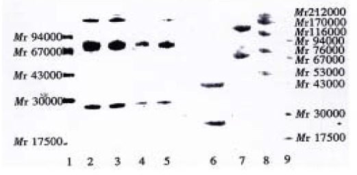
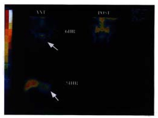
AIM:To determine whether Hb3 and its fragment F(ab')(2) have practical value in radioimmunoimaging of colorectal cancer.METHODS:Intact Hb3 was purified by hydroxylapatite chromatography.The fragment F(ab') (2) was prepared by cold digestion and purified as intact Hb3.Hb3 and its fragment F(ab') (2) were labeled with 99mTc by direct labeling method using SnCl(2) as reducing agent. The radioactive doses ranged from 15 to 40 mCi.The imaging was accomplished by single photon emission computered tomograph (SPECT) with imaging time ranging from 2.5 to 48 hours. In this study, 10 patients were selected. Among them, 7 were administered with intact Hb3, and 3 with F(ab') (2) fragment. All the patients were diagnosed as having colorectal adenocarcinoma.RESULTS:After purification, intact Hb3 and its fragment F(ab') (2) were fit for radioimmunoimaging. The percentage of labeling of (99m)Tc to Hb3 or F(ab') (2) was 80.6%-91.5%. Among the 10 patients, 3 of 7 patients administered with intact Hb3 had positive scans, the other 4 had negative scans, and 2 of 3 patients administered with F(ab') (2)had positive scans, the other 1 had negative scans.CONCLUSION:The results showed that both intact Hb3 and its F(ab') (2) have some practical value in radioimmunoimaging of colorectal cancer, and the effects of imaging with F(ab') (2) was better than that with intact Hb3.
Figures


Similar articles
-
99mTc-Labeled anti-receptor for advanced glycation endproducts monoclonal antibody F(ab’)2 fragments.2010 Jul 16 [updated 2010 Aug 16]. In: Molecular Imaging and Contrast Agent Database (MICAD) [Internet]. Bethesda (MD): National Center for Biotechnology Information (US); 2004–2013. 2010 Jul 16 [updated 2010 Aug 16]. In: Molecular Imaging and Contrast Agent Database (MICAD) [Internet]. Bethesda (MD): National Center for Biotechnology Information (US); 2004–2013. PMID: 20734503 Free Books & Documents. Review.
-
[Preparation of F(ab')2 fragment of monoclonal antibody COC166-9 and its experimental study of radioimmunoimaging for ovarian carcinoma].Zhonghua Fu Chan Ke Za Zhi. 1997 Mar;32(3):152-5. Zhonghua Fu Chan Ke Za Zhi. 1997. PMID: 9596889 Chinese.
-
Comparison of (99m)Tc-labeled PR81 and its F(ab')₂ fragments as radioimmunoscintigraphy agents for breast cancer imaging.Ann Nucl Med. 2011 Feb;25(2):87-92. doi: 10.1007/s12149-010-0434-2. Ann Nucl Med. 2011. PMID: 21061190
-
Noninvasive Evaluation of EGFR Expression of Digestive Tumors Using 99mTc-MAG3-Cet-F(ab')2-Based SPECT/CT Imaging.Mol Imaging. 2022 Jun 24;2022:3748315. doi: 10.1155/2022/3748315. eCollection 2022. Mol Imaging. 2022. PMID: 35903247 Free PMC article.
-
99mTc-Labeled anti-receptor for advanced glycation endproducts polyclonal antibody F(ab’)2 fragments.2010 Jul 16 [updated 2010 Aug 16]. In: Molecular Imaging and Contrast Agent Database (MICAD) [Internet]. Bethesda (MD): National Center for Biotechnology Information (US); 2004–2013. 2010 Jul 16 [updated 2010 Aug 16]. In: Molecular Imaging and Contrast Agent Database (MICAD) [Internet]. Bethesda (MD): National Center for Biotechnology Information (US); 2004–2013. PMID: 20734504 Free Books & Documents. Review.
Cited by
-
Identification of Rho GTPase activating protein 6 isoform 1 variant as a new molecular marker in human colorectal tumors.Pathol Oncol Res. 2010 Sep;16(3):319-26. doi: 10.1007/s12253-009-9226-1. Epub 2009 Dec 4. Pathol Oncol Res. 2010. PMID: 19960375
-
Arterial chemotherapy of 5-fluorouracil and mitomycin C in the treatment of liver metastases of colorectal cancer.World J Gastroenterol. 2002 Aug;8(4):663-7. doi: 10.3748/wjg.v8.i4.663. World J Gastroenterol. 2002. PMID: 12174375 Free PMC article.
-
Preparation and activity of conjugate of monoclonal antibody HAb18 against hepatoma F(ab')(2) fragment and staphylococcal enterotoxin A.World J Gastroenterol. 2001 Apr;7(2):216-21. doi: 10.3748/wjg.v7.i2.216. World J Gastroenterol. 2001. PMID: 11819763 Free PMC article.
-
Early diagnosis for colorectal cancer in China.World J Gastroenterol. 2002 Feb;8(1):21-5. doi: 10.3748/wjg.v8.i1.21. World J Gastroenterol. 2002. PMID: 11833064 Free PMC article. Review.
-
Colorectal carcinoma-associated antigen Ca-Hb3 detected by one-dimensional SDS-polyacrylamide gel electrophoresis and liquid chromatography-tandem mass spectrometry.World J Gastroenterol. 2008 Mar 14;14(10):1588-91. doi: 10.3748/wjg.14.1588. World J Gastroenterol. 2008. PMID: 18330953 Free PMC article.
References
-
- Sun QB, Ho JJI, Kim YS. Human colonic cancer associated antigens detected by three monoclonal antibodies. Chin Med J. 1986;99(1):63–74. - PubMed
-
- Zhang J, Wang CL, Sun QB, Pan AY, Chen SL, Liu CG et al. Radio immunoimaging study with 131I-labeled anti-colorectal carcinoma mono-clonal antibody in nude mouse model. Bull Human Med Univ. 1990;15(3):235–238.
-
- Stanker LH, Vanderlaan M, Juarez-Salinas H. One-step purification of mouse monoclonal antibodies from ascites fluid by hydroxylapatite chromatography. J Immunol Methods. 1985;76:157–169. - PubMed
-
- Ballou B, Reiland J, Levine G, Knowles B, Hakala TR. Tumor location using F(ab')2 mu from a monoclonal IgM antibody: pharmacokinetics. J Nucl Med. 1985;26:283–292. - PubMed
-
- Paik CH, Phan LN, Hong JJ, Sahami MS, Heald SC, Reba RC, Steigman J, Eckelman WC. The labeling of high affinity sites of antibodies with 99mTc. Int J Nucl Med Biol. 1985;12:3–8. - PubMed
LinkOut - more resources
Full Text Sources
