

Dynamic changes of type I,III and IV collagen synthesis and distribution of collagen-producing cells in carbon tetrachloride-induced rat liver fibrosis
- PMID: 11819476
- PMCID: PMC4688608
- DOI: 10.3748/wjg.v5.i5.397
Dynamic changes of type I,III and IV collagen synthesis and distribution of collagen-producing cells in carbon tetrachloride-induced rat liver fibrosis
Abstract
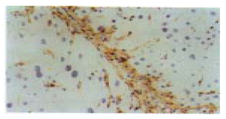
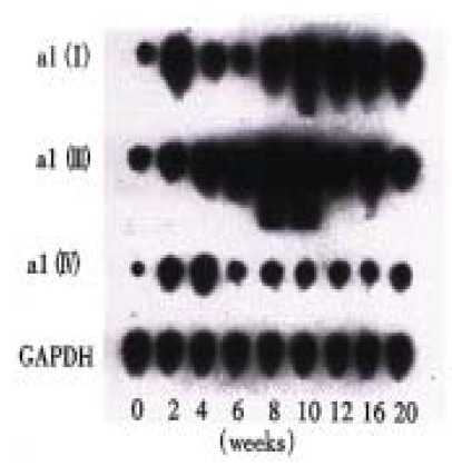
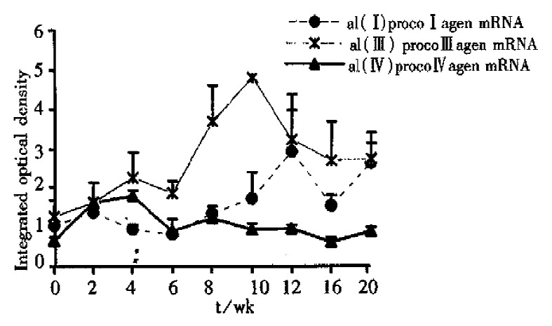
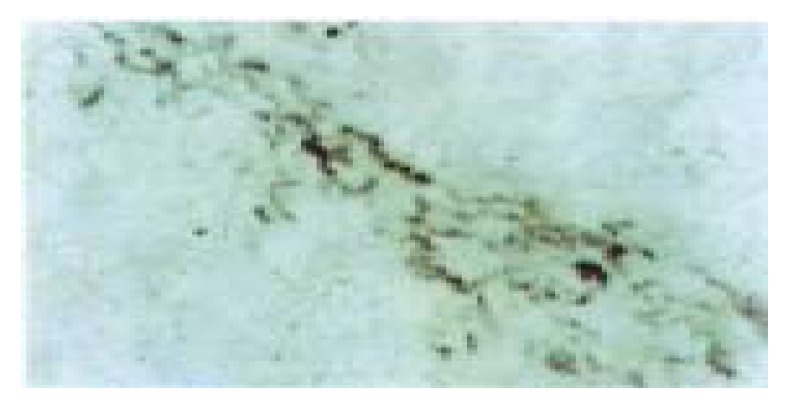
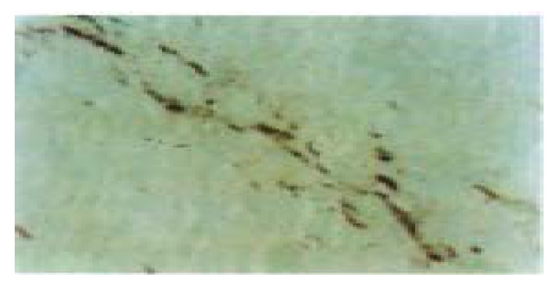
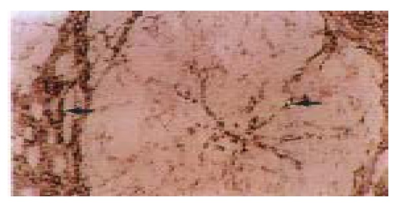
AIM:To find out the relationship between the gene transcription of different types of procollagen and the deposition of the relevant collagens in the liver tissue and to confirm the types of collagen producing cells in liver fibrogenesis.METHODS:Dynamic changes of the expression of alpha1(I), alpha1(III) and alpha1(IV) procollagen mRNA and relevant collagens and the distribution of collagen producing cells during liver fibrogenesis of rat induced by CCl(4) (20 weeks) were investigated with Northern blot analysis, in situ hybridization and immunohistochemical techniques.RESULTS: The increased expression of alpha 1(III) procollagen mRNA by Northern blot analysis was the most predominant one among the three mRNAs during fibrogenesis. However, the enhanced expression of alpha1(IV) procollagen mRNA occurred very early while the expression of alpha1(I) mRNA was not enhanced much until the middle stage of the experiment. Desmin (Dm) positive hepatic stellate cells (HSCs) and few myofibroblasts (MFs) in and around the necrotic areas expressed alpha1(I), alpha1(III) and alpha1(IV) procollagen mRNA signals detected by in situ hybridization at the early stage of the experiment. All the three procollagen mRNA signals thereafter mainly localized in fibroblasts (Fbs) and MFs in fibrotic septa during the middle and late stages of fibrosis, which distributed parallel to the corresponding collagens detected by immunohistochemical study. In addition, the endothelial cells of sinusoids and the small blood vessels within the septa also showed alpha1(IV) procollagen mRNA and type IV collagen expression.CONCLUSION:It is considered that "HSC-MF-Fb effect cell system is the major cellular source of collagen production in liver fibrosis, in which HSCs are collagen producing precursor cells in the early liver fibrogenesis, thereafter the synthesis of type I, III and IV collagens (Col I, Col III and Col IV) mainly derives from MFs and Fbs, which play a very important role in the progress of liver fibrosis. The endothelial cells along sinusoids, as another source of Col IV production, might participate in the capillization of liver sinusoids.
Figures
References
-
- Pierce RA, Glaug MR, Greco RS, Mackenzie JW, Boyd CD, Deak SB. Increased procollagen mRNA levels in carbon tetrachloride-induced liver fibrosis in rats. J Biol Chem. 1987;262:1652–1658. - PubMed
-
- Clement B, Grimaud JA, Campion JP, Deugnier Y, Guillouzo A. Cell types involved in collagen and fibronectin production in normal and fibrotic human liver. Hepatology. 1986;6:225–234. - PubMed
-
- Knittel T, Schuppan D, Meyer zum Büschenfelde KH, Ramadori G. Differential expression of collagen types I, III, and IV by fat-storing (Ito) cells in vitro. Gastroenterology. 1992;102:1724–1735. - PubMed
LinkOut - more resources
Full Text Sources
