

The development of colon innervation in trisomy 16 mice and Hirschsprung's disease
- PMID: 11819726
- PMCID: PMC4688694
- DOI: 10.3748/wjg.v7.i1.16
The development of colon innervation in trisomy 16 mice and Hirschsprung's disease
Abstract
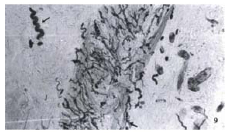
Aim: To study the colon innervation of trisomy 16 mouse, an animal model for Down's syndrome, and the expression of protein gene product 9.5 (PGP 9.5) in the stenosed segment of colon in Hirschsprung's disease (HD).
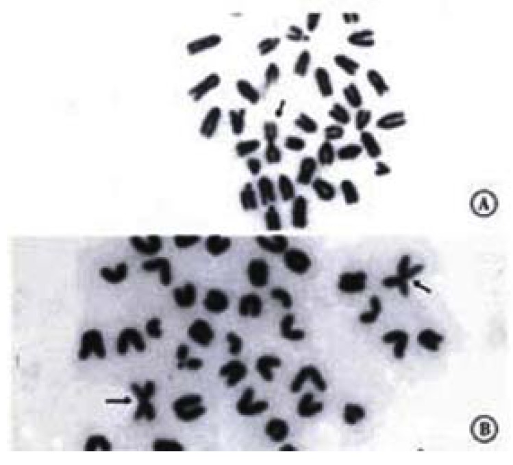
Methods: Trisomy 16 mouse breeding;cytogenetic analysis of trisomy 16 mice; and PG P9.5 immunohistochemistry of colons of trisomy 16 mice and HD were carried out.
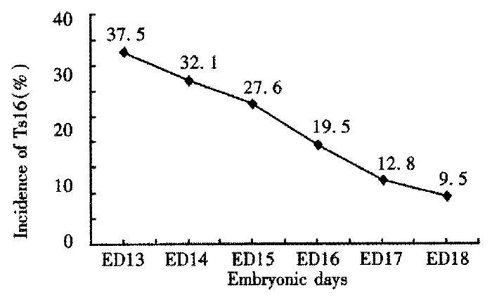
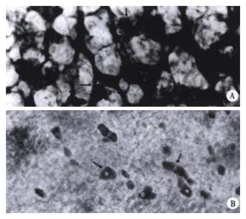
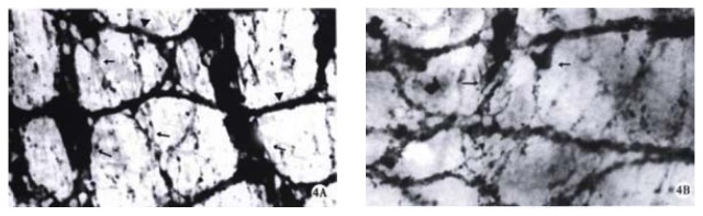
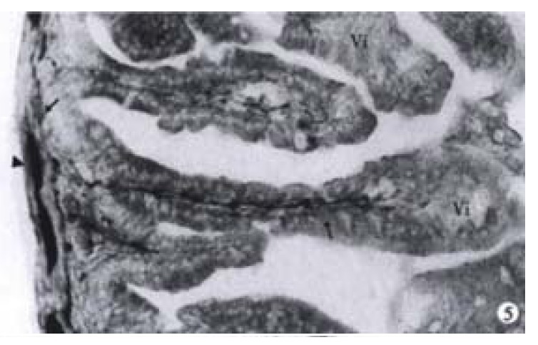
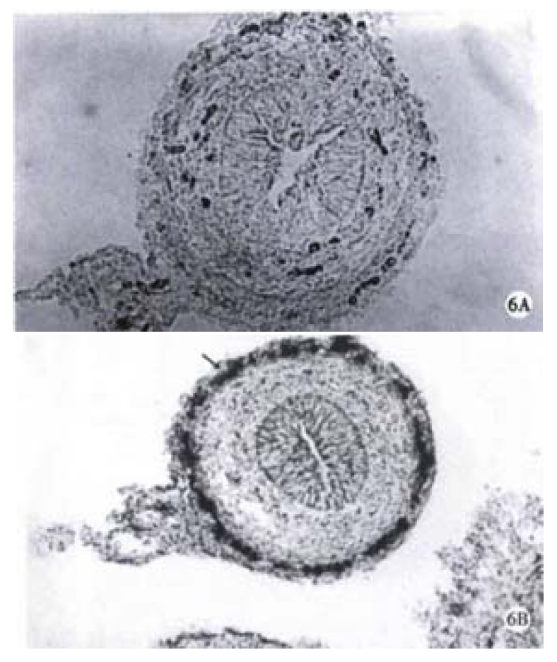
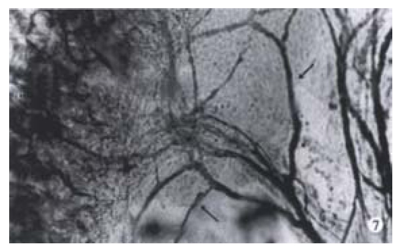
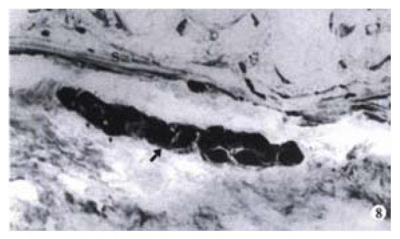
Results: Compared with their normal littermates, the nervous system of colon in trisomy 16 mice was abnormally developed. There existed developmental delay of muscular plexuses of colon, no submucosal plexus was found in the colon, and there was 5mm aganglionic bowel aparting from the anus in trisomy 16 mice. The mesentery nerve fibers were as well developed as shown in their normal littermates. Abundant proliferation of PGP 9.5 positive nerve fibers was revealed in the stenosed segment of HD colon.
Conclusion: Trisomy 16 mice could serve as an animal model for Hirschsprung's disease for aganglionic bowel in the distal part of colon. Abundant proliferation of PGP 9.5 positive fibers resulted from extrinsic nerve compensation, since no ganglionic cells were observed in the stenosed segment of the colon in HD. HD has a genetic tendency.
Figures
References
-
- Li JC, Mi KH, Busch LC, Liu WG, Kühnel W. The expression of protein gene product 9 . 5 in trisomy 16 mouse colon and Hirschsprung's disease. Zhongguo Bingli Shengli Zazhi. 1999;15:1030–1033.
-
- Miyabara S. Animal model for congenital anomalies induced by chromosome aberrations. Cong Anom. 1990;30:49–68.
-
- Epstein CJ, Cox DR, Epstein LB. Mouse trisomy 16: an animal model of human trisomy 21 (Down syndrome) Ann N Y Acad Sci. 1985;450:157–168. - PubMed
-
- Romeo G, Ronchetto P, Luo Y, Barone V, Seri M, Ceccherini I, Pasini B, Bocciardi R, Lerone M, Kääriäinen H. Point mutations affecting the tyrosine kinase domain of the RET proto-oncogene in Hirschsprung's disease. Nature. 1994;367:377–378. - PubMed
-
- Pelet A, Attie T, Goulet O, Eng C, Ponder BA, Munnich A, Lyonnet S. De-novo mutations of the RET proto-oncogene in Hirschsprung's disease. Lancet. 1994;344:1769–1770. - PubMed
Publication types
MeSH terms
Substances
LinkOut - more resources
Full Text Sources
Molecular Biology Databases
Miscellaneous
