

Alterations in metastatic properties of hepatocellular carcinoma cell following H-ras oncogene transfection
- PMID: 11819786
- PMCID: PMC4688718
- DOI: 10.3748/wjg.v7.i3.335
Alterations in metastatic properties of hepatocellular carcinoma cell following H-ras oncogene transfection
Abstract
Aim: To demonstrate the relationship between H-ras oncogene and hepatocellular carcinoma (HCC) metastasis.
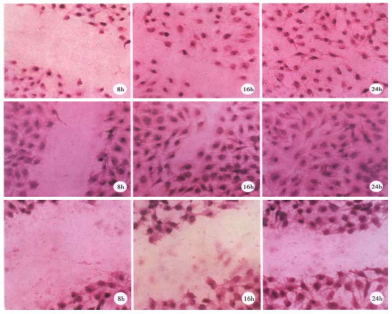
Methods: Activated H-ras oncogene was transfected into SMMC 7721, a cell line derived from human HCC, by calcium phosphate transfection method. Some metastasis-related parameters were detected in vitro, including adhesion assay, migration assay, expression of collagenase IV(c IV ase) and epidermal growth factor receptor (EGFR).
Results: The abilities of H-ras-transfected cell clones in adhesion to laminin (LN) or fibronectin (FN), migration, c IV ase secretion increased markedly, and the expression of EGFR elevated moderately. More importantly, these alterations were consistent positively with the expression of p21, the protein product of H-ras oncogene.
Conclusion: H-ras oncogene could induce the metastatic phenotype of HCC cell in vitro to raise its metastatic potential.
Figures

Similar articles
-
[Alterations in metastatic properties of hepatocellular carcinoma cell following H-ras oncogene transfection].Zhonghua Zhong Liu Za Zhi. 1997 May;19(3):170-2. Zhonghua Zhong Liu Za Zhi. 1997. PMID: 10920888 Chinese.
-
HCRP1 downregulation promotes hepatocellular carcinoma cell migration and invasion through the induction of EGFR activation and epithelial-mesenchymal transition.Biomed Pharmacother. 2017 Apr;88:421-429. doi: 10.1016/j.biopha.2017.01.013. Epub 2017 Jan 22. Biomed Pharmacother. 2017. PMID: 28122307
-
Downregulation of CD147 expression alters cytoskeleton architecture and inhibits gelatinase production and SAPK pathway in human hepatocellular carcinoma cells.J Exp Clin Cancer Res. 2008 Oct 11;27(1):50. doi: 10.1186/1756-9966-27-50. J Exp Clin Cancer Res. 2008. PMID: 18847500 Free PMC article.
-
Alterations of oncogenes, tumor suppressor genes and growth factors in hepatocellular carcinoma: with relation to tumor size and invasiveness.Chin Med J (Engl). 1998 Apr;111(4):313-8. Chin Med J (Engl). 1998. PMID: 10374394
-
The histidine-rich calcium binding protein (HRC) promotes tumor metastasis in hepatocellular carcinoma and is upregulated by SATB1.Oncotarget. 2015 Mar 30;6(9):6811-24. doi: 10.18632/oncotarget.3049. Oncotarget. 2015. PMID: 25762622 Free PMC article.
Cited by
-
Expression of p53 and C-myc genes and its clinical relevance in the hepatocellular carcinomatous and pericarcinomatous tissues.World J Gastroenterol. 2002 Oct;8(5):822-6. doi: 10.3748/wjg.v8.i5.822. World J Gastroenterol. 2002. PMID: 12378623 Free PMC article.
-
Study on the mechanism of epidermal growth factor-induced proliferation of hepatoma cells.World J Gastroenterol. 2003 Feb;9(2):271-5. doi: 10.3748/wjg.v9.i2.271. World J Gastroenterol. 2003. PMID: 12532446 Free PMC article.
-
Combinatorial interventions inhibit TGFβ-driven epithelial-to-mesenchymal transition and support hybrid cellular phenotypes.NPJ Syst Biol Appl. 2015 Nov 26;1:15014. doi: 10.1038/npjsba.2015.14. eCollection 2015. NPJ Syst Biol Appl. 2015. PMID: 28725463 Free PMC article.
-
Expression of liver cancer associated gene HCCA3.World J Gastroenterol. 2001 Dec;7(6):821-5. doi: 10.3748/wjg.v7.i6.821. World J Gastroenterol. 2001. PMID: 11854909 Free PMC article.
-
K-ras gene mutation in the diagnosis of ultrasound guided fine-needle biopsy of pancreatic masses.World J Gastroenterol. 2003 Jan;9(1):188-91. doi: 10.3748/wjg.v9.i1.188. World J Gastroenterol. 2003. PMID: 12508380 Free PMC article.
References
-
- Tang ZY. Advances in clinical research of hepatocellular carcinoma in China. World J Gastroenterol. 1998;4(Suppl 2):4–7.
MeSH terms
Substances
LinkOut - more resources
Full Text Sources
Medical
Research Materials
Miscellaneous
