

Review
doi: 10.1136/bmj.324.7332.283.
Childhood leukaemia
Affiliations
- PMID: 11823363
- PMCID: PMC1122200
- DOI: 10.1136/bmj.324.7332.283
Item in Clipboard
Review
Childhood leukaemia
BMJ.
.
No abstract available
Figures
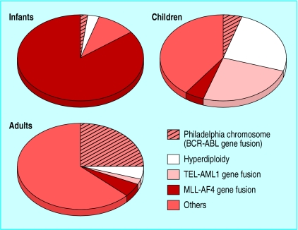
Major molecular subsets of acute lymphoblastic leukaemia in infants (<1 year old), children (2-10 years old), and adults
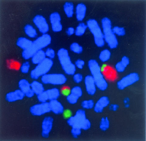
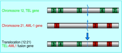
Chromosomal translocation to form the TEL-AML1 fusion gene in childhood acute lymphoblastic leukaemia. Top: Fluorescence in situ hybridisation labelling of dividing leukaemic cell chromosomes with specific probes for chromosome 12 (red) and chromosome 21 (green) reveal two red and green chromosomes (one large, one small). These are copies of chromosomes 12 and 21 between which there has been a reciprocal exchange of DNA. Bottom: The TEL and AML1 genes lie at the breaks and are brought together by the exchange. The genes break in non-coding (grey) regions between the coding regions (numbered, green or red), and re-joining of the two broken genes forms a novel fusion gene
Chromosomal translocation to form the TEL-AML1 fusion gene in childhood acute lymphoblastic leukaemia. Top: Fluorescence in situ hybridisation labelling of dividing leukaemic cell chromosomes with specific probes for chromosome 12 (red) and chromosome 21 (green) reveal two red and green chromosomes (one large, one small). These are copies of chromosomes 12 and 21 between which there has been a reciprocal exchange of DNA. Bottom: The TEL and AML1 genes lie at the breaks and are brought together by the exchange. The genes break in non-coding (grey) regions between the coding regions (numbered, green or red), and re-joining of the two broken genes forms a novel fusion gene
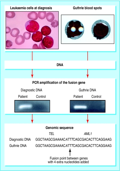
Identification of fusion genes in neonatal blood spots of patients with leukaemia. At diagnosis of childhood acute lymphoblastic leukaemia, a TEL-AML1 fusion gene can be identified in the leukaemic cells. The TEL-AML1 sequence is first determined by long range PCR, then oligonucleotide primers are designed for that unique sequence and for use in short range (conventional) PCR. DNA is extracted from a diagnostic sample for PCR and, in parallel, a segment from a neonatal blood spot is subjected to PCR. If successful, both samples from the patient amplify to produce a nucleotide sequence visualised as a band in a gel. Sequencing of these bands shows them to be identical
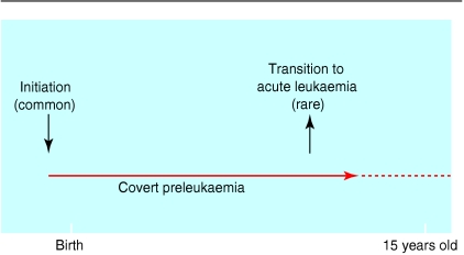
Natural course of childhood leukaemia
References
-
- Rowley JD. The critical role of chromosome translocations in human leukemias. Annu Rev Genet. 1998;32:495–519. - PubMed
-
- Kersey JH. Fifty years of studies of the biology and therapy of childhood leukemia. Blood. 1997;90:4243–4251. - PubMed
-
- Greaves M. Molecular genetics, natural history and the demise of childhood leukaemia. Eur J Cancer. 1999;35:173–185. - PubMed
-
- Greaves M. Cancer. The evolutionary legacy. Oxford: Oxford University Press; 2000.
-
- Ford AM, Ridge SA, Cabrera ME, Mahmoud H, Steel CM, Chan LC, et al. In utero rearrangements in the trithorax-related oncogene in infant leukaemias. Nature. 1993;363:358–360. - PubMed
Publication types
MeSH terms
Substances
LinkOut - more resources
Full Text Sources
Medical