. 2002 Feb 1;21(3):202-10.
doi: 10.1093/emboj/21.3.202.

Post-natal knockout of prion protein alters hippocampal CA1 properties, but does not result in neurodegeneration

Affiliations

Post-natal knockout of prion protein alters hippocampal CA1 properties, but does not result in neurodegeneration

G R Mallucci et al. EMBO J. .

Erratum in

  • EMBO J 2002 Mar 1;21(5):1240

Abstract

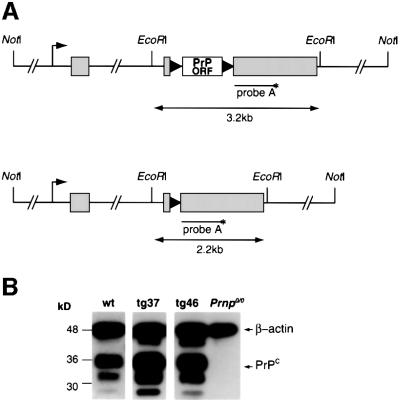
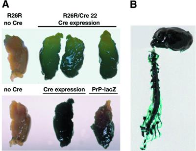
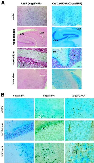
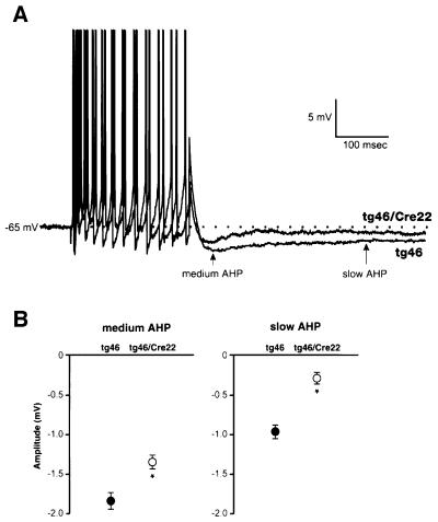
Prion protein (PrP) plays a crucial role in prion disease, but its physiological function remains unclear. Mice with gene deletions restricted to the coding region of PrP have only minor phenotypic deficits, but are resistant to prion disease. We generated double transgenic mice using the Cre-loxP system to examine the effects of PrP depletion on neuronal survival and function in adult brain. Cre-mediated ablation of PrP in neurons occurred after 9 weeks. We found that the mice remained healthy without evidence of neurodegeneration or other histopathological changes for up to 15 months post-knockout. However, on neurophysiological evaluation, they showed significant reduction of afterhyperpolarization potentials (AHPs) in hippocampal CA1 cells, suggesting a direct role for PrP in the modulation of neuronal excitability. These data provide new insights into PrP function. Furthermore, they show that acute depletion of PrP does not affect neuronal survival in this model, ruling out loss of PrP function as a pathogenic mechanism in prion disease and validating therapeutic approaches targeting PrP.


Figures

None
Fig. 1. The MloxP construct and PrP expression in tg37 and tg46 mice. (A) MloxP construct containing the floxed murine PrP coding region (PrP ORF, white box) within the CosSHaTet expression vector (exons represented by grey boxes) before (upper image) and after Cre-mediated recombination (lower image). Black triangles represent loxP sequences. The annealing site of DNA probe A used in Southern blotting is shown. (B) Western blotting showing PrP expression in wild-type, tg37 and tg46 mouse brains using antibody 6H4 (Prionics). β-actin was simultaneously detected to control for loading differences between the samples. After quantitation of relative signal intensities, expression levels of PrP by tg46 and wild-type mice are similar and ∼3-fold lower than in tg37 mice.
None
Fig. 2. NFH–Cre transgene, temporal expression. (A) The NFH–Cre construct containing the Cre-coding region (grey box) inserted into exon 1 (E1) of the murine NFH gene upstream of the NFH start codon (ATG). Black box, SV40 polyadenylation signal; white boxes, NFH exons. NLS/ATG, nuclear localization signal and start codon of Cre gene. (B) Southern blotting of EcoRI-digested DNA from the brains of two tg46–Cre 22 (lanes 1 and 2), four tg46 (lanes 4–6 and 11) and two tg37–Cre 22 (lanes 8 and 9) mice. DNA from a Prnp0/0 mouse brain was loaded in lane 10. Hybridization of probe with unrecombined 3.2 kb and recombined 2.2 kb fragments is seen in double transgenic animals, the proportions of each fragment are shown beneath the image. There is no recombination in tg46 mice. (C) A histogram showing the time course of onset of Cre-mediated recombination of the MloxP locus. DNA from brains from three or more tg46–Cre 22 or tg37–Cre 22 mice or embryos were analysed for recombination by Southern blotting as above. E, embryonic day; P, post-natal day; w, weeks of age. Mean values for the number of mice indicated are depicted. For all time points up to 12 weeks, n = 3.
None
Fig. 3. Spatial features of NFH–Cre transgene expression in R26R–Cre 22 mice at the macroscopic level. (A) LacZ expression in R26R–Cre 22 mice and their R26R littermates (top panel) was determined by x-gal staining of brains. There is strong lacZ expression in whole brains of R26R–Cre 22 mice where Cre expression has allowed transcription of the lacZ gene. There is no lacZ expression in the absence of Cre, in the R26R brain. In the lower panel, a lacZ-expressing transgenic mouse, PrP-lacZ, which has high forebrain expression of lacZ (E.A.Asante, unpublished data), was used as a positive control; the R26R–Cre 22 mouse brain (centre) shows lacZ expression throughout fore and hind brain in comparison. (B) LacZ expression was seen throughout the central and peripheral nervous system in R26R–Cre 22 mice.
None
Fig. 4. Spatial features of NFH–Cre transgene expression in R26R–Cre 22 mice at the histological level. (A) The x-gal-stained brains of the R26R–Cre 22 and R26R mice above were sectioned and counterstained with Nuclear Fast Red (NFR). The strong expression of lacZ was seen throughout the brains of R26R–Cre 22 mice, particularly in neuron-rich regions such as the hippocampus. No lacZ expression was detected in the absence of Cre expression in R26R brains. (B) Sections from these mice were counter-immunostained using antibodies against NFH and GFAP to define cell types expressing lacZ. x-gal and NFH staining are superimposed (centre panel), whereas GFAP staining is distinct from the x-gal-stained cells and regions, confirming neuronal, but not astrocytic, distribution of Cre expression. Scale bar = 100 µm. CA1 and CA3 regions of hippocampus are indicated. An astrocyte is boxed. Arrows indicate pyramidal cells in cerebellum. mol, molecular layer; gr, granule cell layer of cerebellum.
None
Fig. 5. Knockout of PrP expression in brains of tg46–Cre 22 mice. (A) Immunohistochemistry for PrPc in wild-type, tg46, tg46–Cre 22 and Prnp0/0 brains. Loss of PrP immunoreactivity to ICSM18 was seen in tg46–Cre 22 mouse brain sections, producing appearances equivalent to ICSM15-stained sections, which did not bind to mouse brain proteins and acted as a negative control. Serial frozen sections were stained for PrPc with ICSM18 and 6H4 and with anti-gangliocerebrosidase C (α-G) and ICSM15 antibodies. Counterstaining was with haematoxylin. (B) Macroscopic appearance of sections from tg46 and tg46–Cre 22 mouse brains immunostained as above. The staining patterns with α-G (positive control) and ICSM15 (negative control) were equivalent in the tg46 and conditional knockout mice, but the signal from PrP was undetectable with both 6H4 and ICSM18 in tg46–Cre 22 brain. (C) Section of tg46–Cre 22 mouse containing choroid plexus (CP) shows immunostaining of ependymal cells by 6H4 antibody, which does not occur in Prnp0/0 sections. Scale bar = 100 µm.
None
Fig. 6. Abolition of AHP in hippocampal CA1 cells of conditional PrP-knockout mice. (A) Sample traces showing the AHPs following a burst of 12 action potentials in CA1 pyramidal cells (action potentials have been truncated). In tg46–Cre 22, amplitudes of both the medium and slow AHPs were significantly reduced compared with tg46. (B) Average medium and slow AHPs in the tg46 and tg46–Cre 22 groups. Results extracted from ANOVA test. Values were derived from all numbers of action potentials; SEMs represent the variance after that attributable to the different numbers of action potentials had been accounted for and removed (*two–way ANOVA, P <0.001 for both medium and slow AHPs; tg46, n = 10 cells; tg46–Cre 22, n = 14 cells).

References

    1. Barrow P.A., Holmgren,C.D., Tapper,A.J. and Jeffreys,J.G.R. (1999) Intrinsic physiological and morphological properties of principal cells of the hippocampus and neocortex in hamsters infected with scrapie. Neurobiol. Dis., 6, 406–423. - PubMed
    1. Barrow P.A., Mallucci,G.R., Collinge,J. and Jefferys,J.G.R. (2000) Rescue of a physiological phenotype in mice with ablated PrP gene but expressing a transgene cassette for PrP. J. Physiol., 523P, 203P (Abstract).
    1. Brandner S., Isenmann,S., Raeber,A., Fischer,M., Sailer,A., Kobayashi,Y., Marino,S., Weissmann,C. and Aguzzi,A. (1996) Normal host prion protein necessary for scrapie-induced neurotoxicity. Nature, 379, 339–343. - PubMed
    1. Brown D.R. et al. (1997a) The cellular prion protein binds copper in vivo. Nature, 390, 684–687. - PubMed
    1. Brown D.R., Schulz-Schaeffer,W.J., Schmidt,B. and Kretzschmar,H.A. (1997b) Prion protein-deficient cells show altered response to oxidative stress due to decreased SOD-1 activity. Exp. Neurol., 146, 104–112. - PubMed