. 2002 Feb 1;21(3):231-9.
doi: 10.1093/emboj/21.3.231.

The crystal structure of the zymogen catalytic domain of complement protease C1r reveals that a disruptive mechanical stress is required to trigger activation of the C1 complex

Affiliations

The crystal structure of the zymogen catalytic domain of complement protease C1r reveals that a disruptive mechanical stress is required to trigger activation of the C1 complex

Monika Budayova-Spano et al. EMBO J. .

Abstract

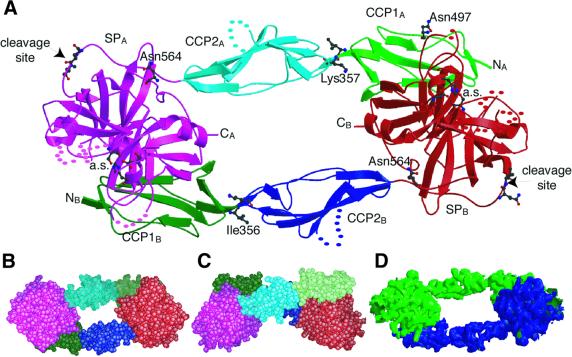
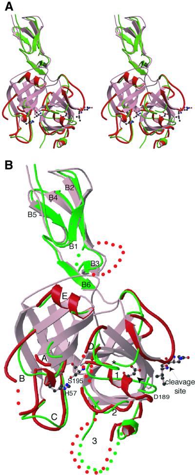
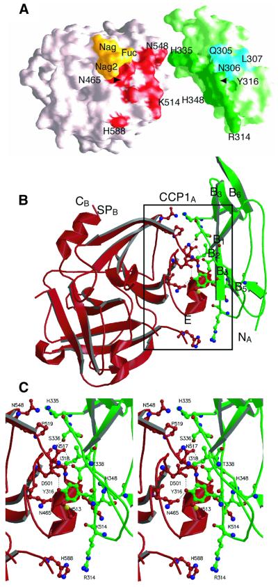
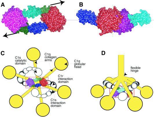
C1r is the modular serine protease (SP) that mediates autolytic activation of C1, the macromolecular complex that triggers the classical pathway of complement. The crystal structure of a mutated, proenzyme form of the catalytic domain of human C1r, comprising the first and second complement control protein modules (CCP1, CCP2) and the SP domain has been solved and refined to 2.9 A resolution. The domain associates as a homodimer with an elongated head-to-tail structure featuring a central opening and involving interactions between the CCP1 module of one monomer and the SP domain of its counterpart. Consequently, the catalytic site of one monomer and the cleavage site of the other are located at opposite ends of the dimer. The structure reveals unusual features in the SP domain and provides strong support for the hypothesis that C1r activation in C1 is triggered by a mechanical stress caused by target recognition that disrupts the CCP1-SP interfaces and allows formation of transient states involving important conformational changes.


Figures

None
Fig. 1. Homodimeric structure of the CCP1–CCP2–SP C1r catalytic domain. (A) Overall view of the structure of the zymogen. CCP1 modules are in green, CCP2 modules in blue and SP domains in magenta (molecule A) or red (molecule B). The residues at the catalytic sites (a.s.) and at the cleavage sites are shown, as are the residues Asn497 and Asn564, which bear oligosaccharide chains, and the residues Ile356 and Lys357 at the CCP1–CCP2 interface. NA, NB and CA, CB indicate the N- and C-terminal ends of molecules A and B. Dots represent disordered segments. (B and C) Space-filling representations of the bottom and side views of the structure. (D) Electron density map of the activated domain at 4 Å resolution. The map was contoured at the 1σ level and smoothed by solvent flattening (see Materials and methods).
None
Fig. 2. Three-dimensional structure of the CCP2–SP region of C1r and comparison with the homologous region of C1s: (A) stereoview; (B) detailed representation of the structures. The structures of C1r (red) and C1s (green) are superimposed. The loops in the SP domains are labeled according to Perona and Craik (1997), and the β-strands in the CCP module are numbered from B1 to B6. In the C1r structure, the His485(57) and Ser637(195) residues of the catalytic triad, the mutated Gln446(15)–Ile447(16) cleavage site, and Asp631(189) are shown with ball and sticks. The asterisk indicates the position of Ile423(16) in C1s after activation. Dots represent disordered segments. In the SP domain of C1s, only surface loops differing from the common core are shown for the sake of clarity.
None
Fig. 3. Structural alignments of (A) the SP domains of C1r, C1s and chymotrypsinogen, and (B) the CCP modules of C1r, C1s and complement receptor 2. The residue numberings of C1r and chymotrypsinogen are shown in (A). In (B), the residues with a β-strand conformation are underlined, and the strand numbering is indicated above. The residues involved in the major CCP1–SP interaction in C1r, and in the interaction between the CCP2 module of CR2 and its C3d ligand, are indicated by asterisks. The residues involved in the additional interaction observed only at the CCP1A–SPB interface are indicated by a ‘#’. Hyp_V, hypervariable loop.
None
Fig. 4. Structure at the CCP1A–SPB interface. (A) Surface representation illustrating shape complementarity between SPB (left) and CCP1A (right). CCP1A was shifted and rotated to the right for clarity. Areas involved in the major interaction common to both CCP1–SP interfaces are shown in red (SPB) and dark green (CCP1A). The areas involved in the additional interaction observed only at the CCP1A–SPB interface are colored yellow (SPB) and blue (CCP1A). (B) Overall structure of the assembly between SPB (red) and CCP1A (green). (C) Stereoview of the CCP1A–SPB interface. Only the major interaction is shown for clarity. The hydrogen bonding network at the interface is depicted by dotted lines. Strand B4 and the N-terminal part of strand B2 are not shown in ribbon representation for the sake of clarity.
None
Fig. 5. Functional implications of the dimeric structure of the C1r catalytic domain in the context of the C1 complex. (A) Resting head-to-tail configuration of the C1r catalytic domain. Arrows illustrate the triggering stress required to achieve the transient conformational state needed for activation of an SP domain by its counterpart (B). (C and D) The C1r catalytic domain in the context of the C1 complex: (C) bottom and (D) side views of a macroscopic model of C1 (modified from Arlaud et al., 1987b).

References

    1. Arlaud G.J., Gagnon,J., Villiers,C.L. and Colomb,M.G. (1986) Molecular characterization of the catalytic domains of human complement serine protease C1r. Biochemistry, 25, 5177–5182. - PubMed
    1. Arlaud G.J., Willis,A.C. and Gagnon,J. (1987a) Complete amino acid sequence of the A chain of human complement-classical-pathway enzyme C1r. Biochem. J., 241, 711–720. - PMC - PubMed
    1. Arlaud G.J., Colomb,M.G. and Gagnon,J. (1987b) A functional model of the human C1 complex. Immunol. Today, 8, 106–111. - PubMed
    1. Arlaud G.J., Volanakis,J.E., Thielens,N.M., Narayana,S.V.L., Rossi,V. and Xu,Y. (1998) The atypical serine proteases of the complement system. Adv. Immunol., 69, 249–307. - PubMed
    1. Bernstein F.C., Koetzle,T.F., Williams,G.J., Meyer,E.E.,Jr, Brice,M.D., Rodgers,J.R., Kennard,O., Shimanouchi,T. and Tasumi,M. (1977) The Protein Data Bank: a computer-based archival file for macromolecular structures. J. Mol. Biol., 112, 535–542. - PubMed

Publication types