. 2002 Feb 1;21(3):251-8.
doi: 10.1093/emboj/21.3.251.

Contrasting requirements for ubiquitylation during Fc receptor-mediated endocytosis and phagocytosis

Affiliations

Contrasting requirements for ubiquitylation during Fc receptor-mediated endocytosis and phagocytosis

James W Booth et al. EMBO J. .

Abstract

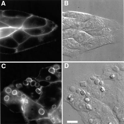
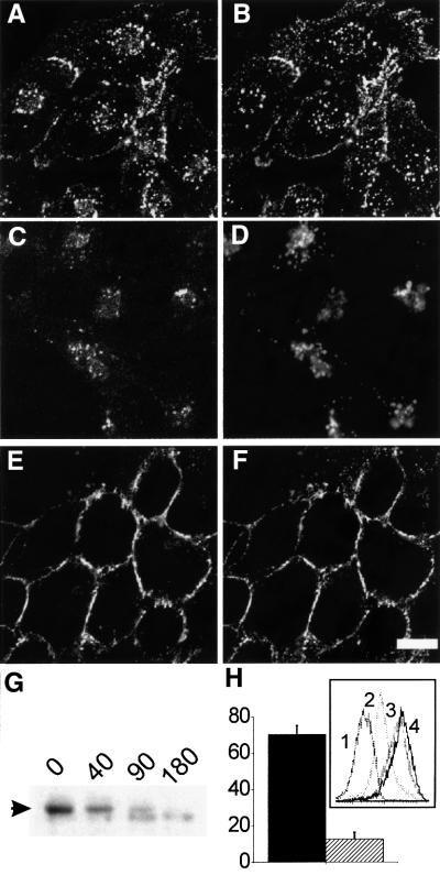
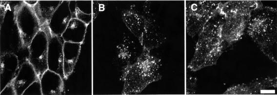
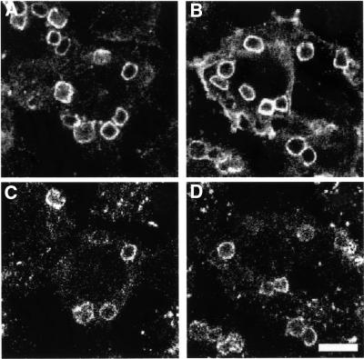
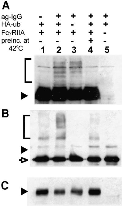
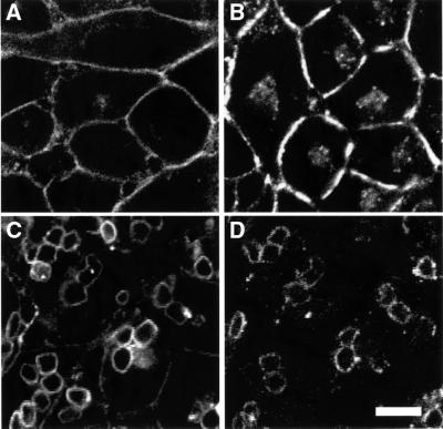
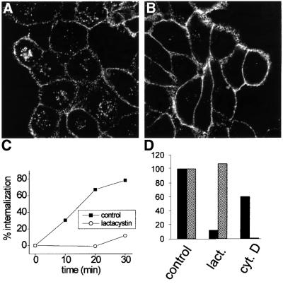
Fc receptors on leukocytes mediate internalization of antibody-containing complexes. Soluble immune complexes are taken up by endocytosis, while large antibody-opsonized particles are internalized by phagocytosis. We investigated the role of ubiquitylation in internalization of the human FcgammaRIIA receptor by endocytosis and phagocytosis. A fusion of FcgammaRIIA to green fluorescent protein (GFP) was expressed in ts20 cells, which bear a temperature-sensitive mutation in the E1 ubiquitin-activating enzyme. Uptake of soluble IgG complexes mediated by FcgammaRIIA-GFP was blocked by incubation at the restrictive temperature, indicating that endocytosis requires ubiquitylation. In contrast, phagocytosis and phagosomal maturation were largely unaffected when ubiquitylation was impaired. FcgammaRIIA-GFP was ubiquitylated in response to receptor cross-linking. Elimination of the lysine residues present in the cytoplasmic domain of FcgammaRIIA impaired endocytosis, but not phagocytosis. The proteasomal inhibitor clasto-lactacystin beta-lactone strongly inhibited endocytosis, but did not affect phagocytosis. These studies demonstrate a role for ubiquitylation in the endocytosis of immune receptors, and reveal fundamental differences in the mechanisms underlying internalization of a single receptor depending on the size or multiplicity of the ligand complex.


Figures

None
Fig. 1. FcγRIIA–GFP mediates phagocytosis in ts20 cells. GFP fluorescence was visualized in living ts20 cells expressing FcγRIIA–GFP. (A) Fluorescence of cells with no addition of particles. (C) Fluorescence of cells incubated with IgG-coated fixed SRBCs for 30 min at 37°C. (B and D) Corresponding DIC images. Bar = 8 µm.
None
Fig. 2. Endocytosis of FcγRIIA–GFP in ts20 cells is blocked by temperature shift. ts20 cells expressing FcγRIIA–GFP were incubated with ag-IgG without (A–D) or with (E and F) prior incubation for 1.75 h at 42°C. (A, C and E) Distribution of FcγRIIA–GFP. (B and F) Distribution of ag-IgG detected with Cy3-anti-human antibody. (D) Distribution of LAMP-1 by immunofluorescence in the same cells as (C). Incubation with ag-IgG was at 37°C for 40 min (A, B, E and F) or 80 min (C and D). Bar = 8 µm. (G) ts20 cells expressing FcγRIIA–GFP were incubated with ag-IgG for 0, 40, 90 or 180 min, then total cell lysates were prepared and analyzed by western blotting with anti-GFP antibodies. The arrow marks the position of FcγRIIA–GFP; the lower band is a non-specific background band. (H) Cells were pre-incubated at 37°C (solid bar) or 42°C (hatched bar) for 1–1.75 h, then endocytosis was analyzed by flow cytometry. Bars indicate internalization of cross-linking antibody as a percentage of initial amount bound (n = 4 experiments, ±SE). Inset: Cy5 fluorescence curves from an individual experiment. Abscissa: relative fluorescence intensity (log scale). Ordinate: number of cells. Curve 1, background (no anti-FcγRII antibody); curve 2, post-cross-linking, cells pre-incubated at 37°C; curve 3, post-cross-linking, cells pre-incubated at 42°C; curve 4, no internalization (cross-linking at 4°C).
None
Fig. 3. Endocytosis of FcγRIIA–GFP in E36 cells. (A) Fluorescence of E36 cells expressing FcγRIIA–GFP prior to addition of ag-IgG. (B and C) Cells were incubated with ag-IgG for 40 min without (B) or with (C) pre-incubation for 1.75 h at 42°C. Representative of five experiments. Bar = 8 µm.
None
Fig. 4. Phagocytosis via FcγRIIA–GFP in ts20 cells is not blocked by temperature shift. ts20 cells expressing FcγRIIA–GFP were incubated for 45 min with opsonized SRBCs after pre-incubation for 1.75 h at 37°C (A and C) or 42°C (B and D). (A and B) Distribution of FcγRIIA–GFP. (C and D) Localization of LAMP-1 in corresponding cells. Bar = 8 µm.
None
Fig. 5. FcγRIIA–GFP is ubiquitylated upon receptor aggregation. (A) Total cell lysates were analyzed by SDS–PAGE and immunoblotting with polyclonal anti-GFP antibody. Lane 2: cells stably transfected with FcγRIIA–GFP and transiently transfected with HA-ubiquitin were incubated with ag-IgG at 37°C for 10 min before preparation of lysates. Conditions for other lanes were the same as for lane 2, with the following exceptions: lane 1, no addition of ag-IgG; lane 3, no transfection with HA-ubiquitin; lane 4, cells were pre-incubated at 42°C for 1.75 h before addition of ag-IgG; lane 5, no transfection with FcγRIIA–GFP. (B) FcγRIIA–GFP was immunoprecipitated from the lysates used in (A) with mouse anti-GFP and was analyzed by SDS–PAGE and immunoblotting with anti-HA. Lanes correspond to those in (A). (C) The blot from (B) was reprobed with rabbit anti-GFP antibody to compare total amounts of receptor loaded. The position of the 66 kDa marker is indicated by a filled arrowhead on each blot. The brackets indicate the region in which polyubiquitylated forms of FcγRIIA–GFP migrate. The open arrow indicates the immunoprecipitating antibody. The band seen at the position of the 66 kDa marker in (B) is present in all lanes and thus reflects non-specific binding of antibodies.
None
Fig. 6. FcγRIIA/5KR is deficient in endocytosis, but not in phagocytosis. (A) Fluorescence of ts20 cells expressing FcγRIIA/5KR–GFP. (B and C) Fluorescence after incubation for 40 min at 37°C with ag-IgG (B) or IgG-coated fixed SRBCs (C). Images in (A) and (B) were acquired with identical gain settings on the confocal microscope. (D) Distribution of LAMP-1 in the same cells as (C). Images reflect at least four experiments. Bar = 8 µm.
None
Fig. 7. Inhibition of the proteasome impairs endocytosis, but not phagocytosis. (A and B) ts20 cells expressing FcγRIIA-MH were incubated with DMSO vehicle (A) or 20 µM clasto-lactacystin β-lactone (B) for 1 h, then ag-IgG was added for 40 min to induce endocytosis. Ag-IgG was detected with Cy3-anti-human secondary antibody after cell permeabilization. (C) The kinetics of endocytosis of immune complexes via FcγRIIA–GFP were analyzed by flow cytometry in cells pre-treated for 1 h with DMSO (control; solid squares) or 20 µM clasto-lactacystin β-lactone (open circles). Values are the means of two experiments. (D) Cells were pre-incubated with 20 µM clasto-lactacystin β-lactone (lact.) for 1 h or with 10 µM cytochalasin D (cyt. D) for 10 min. Inhibitors were also present during the uptake assays, except for clasto-lactacystin β-lactone during endocytosis. Endocytosis (solid bars) and phagocytosis (hatched bars) were assayed for 30 min at 37°C by flow cytometry or by scoring phagocytic index. Levels are expressed as a percentage of control (DMSO vehicle alone) and reflect means of two experiments.

References

    1. Aggeler J. and Werb,Z. (1982) Initial events during phagocytosis by macrophages viewed from outside and inside the cell: membrane–particle interactions and clathrin. J. Cell Biol., 94, 613–623. - PMC - PubMed
    1. Cenciarelli C., Wilhelm,K.G.,Jr, Guo,A. and Weissman,A.M. (1996) T cell antigen receptor ubiquitination is a consequence of receptor-mediated tyrosine kinase activation. J. Biol. Chem., 271, 8709–8713. - PubMed
    1. Daeron M. (1997) Fc receptor biology. Annu. Rev. Immunol., 15, 203–234. - PubMed
    1. Downey G.P., Botelho,R.J., Butler,J.R., Moltyaner,Y., Chien,P., Schreiber,A.D. and Grinstein,S. (1999) Phagosomal maturation, acidification and inhibition of bacterial growth in nonphagocytic cells transfected with FcγRIIA receptors. J. Biol. Chem., 274, 28436–28444. - PubMed
    1. Duchemin A.M., Ernst,L.K. and Anderson,C.L. (1994) Clustering of the high affinity Fc receptor for immunoglobulin G (FcγRI) results in phosphorylation of its associated γ-chain. J. Biol. Chem., 269, 12111–12117. - PubMed

Publication types