. 2002 Feb 1;21(3):440-50.
doi: 10.1093/emboj/21.3.440.

Transvection effects involving DNA methylation during meiosis in the mouse

Affiliations

Transvection effects involving DNA methylation during meiosis in the mouse

Minoo Rassoulzadegan et al. EMBO J. .

Abstract

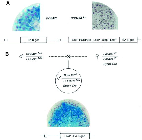
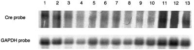
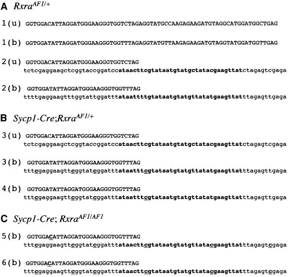
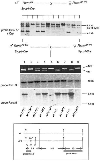
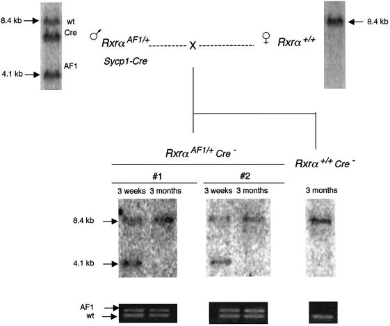
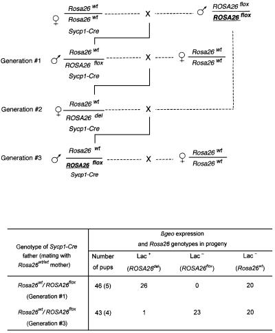
High efficiencies of recombination between LoxP elements were initially recorded when the Cre recombinase was expressed in meiotic spermatocytes. However, it was unexpectedly found that LoxP recombination fell to very low values at the second generation of mice expressing Cre during meiosis. The inability of the LoxP elements to serve as recombination substrates was correlated with cytosine methylation, initially in LoxP and transgene sequences, but later extending for distances of at least several kilobases into chromosomal sequences. It also affected the allelic locus, implying a transfer of structural information between alleles similar to the transvection phenomenon described in Drosophila. Once initiated following Cre-LoxP interaction, neither cis-extension nor transvection of the methylated state required the continuous expression of Cre, as they occurred both in germinal and somatic cells and in the fraction of the offspring that had not inherited the Sycp1-Cre transgene. Therefore, these processes depend on a physiological mechanism of establishment and extension of an epigenetic state, for which they provide an experimental model.


Figures

None
Fig. 1. Efficient excision of a floxed cassette in the testis of a male who inherited the Sycp1-Cre transgene. (A) Left: x-gal staining of a section of an adult (4 months) ROSA26 male mouse showing β-galactosidase expression at all stages of germinal differentiation. Right: no expression is detectable in a male of the same age carrying the floxed locus (ROSA26flox). (B) The indicated cross generated heterozygous ROSA26flox/Rosa26wt;Sycp1-Cre males. x-gal staining shows extensive β-galactosidase accumulation in testicular germ cells starting at the pachytene stage (note that the earliest stages at the periphery of the tubule remain negative), demonstrating excision of the floxed cassette generating the ROSA26del recombined allele.
None
Fig. 2. The floxed cassette is not excised after propagation for more than one generation of the Sycp1-Cre transgene. Same experiment as in Figure 1, but the floxed ROSA26 locus was transferred to a third generation progeny in one of the Sycp1-Cre;ROSA26 families. Indicated in the table are the numbers of β-galactosidase-expressing mice in the male progeny (ROSA26del) and, among the negative phenotypes, the wild-type and floxed genotypes distinguished by PCR analysis. Note that meiosis in the male, indicated as Generation #1, was the first exposure of the floxed locus to the recombinase. The total number of pups analyzed is indicated, with the number of litters shown in parentheses.
None
Fig. 3. Expression of the Cre transgene in males that have not excised the floxed cassette. Northern blot analysis of testis RNA of males designated as generation #1 (lanes 1–6) and generation #3 (lanes 7–13) in the experiment described in Figure 2. Hybridization was performed with a Cre probe amplified from pBS185 DNA with primers Cre1 and Cre2 and a GAPDH probe as a control (Lopez et al., 1999).
None
Fig. 4. Cytosine methylation in the LoxP elements of the ROSA26flox locus. Bisulfite treatment and subsequent PCR amplification and sequencing (Olek et al., 1996) were performed to determine the extent of cytosine methylation. Methylated cytosines are detected as cytosines (indicated as bold underlined characters) and the unmethylated bases as thymine nucleotides. u, untreated DNA; b, bisulfite-treated DNA. Sequence 1 shows amplification from testis DNA of a ROSA26 mouse without the Cre transgene (sequences identical in 12 amplified clones per animal and in two animals); sequences 2.1 and 2.2 are different sequences obtained from the ROSA26del testis of Rosa26wt/ROSA26flox male (generation #1 in Figure 2, the two sequences being found among 12 amplified clones for three animals of the same generation), and sequences 3 and 4 show amplification from the somatic (tail) DNA of two groups of three males (six strands sequenced for each animal all showing the same pattern). Amplification was performed with primers Rosa26–1S, 2S, 1R and Rosa26–1S, 2S, ch3R (see Materials and methods).
None
Fig. 5. Cytosine methylation of LoxP inhibits Cre recombination. Actively growing STO cells were transfected with a mixture of 1 µg of pBS185 (Cre expression vector) and 0.2 µg of pβgeo–LoxP plasmid DNA. DNA was prepared 72 h after transfection. PCR amplification of a 560 bp fragment with primers Neo1 and Neo2 (1 min at 94°C, 1 min at 60°C, 15 s at 72°C, 25 cycles) was performed as a control on undiluted extracts. PCR analysis with primers PBS1 and PBS2 (1 min at 94°C, 1 min at 56°C, 15 s at 72°C, 30 cycles) was then performed to amplify a fragment of 81 bp diagnostic of the excision of the floxed βgeo region, revealed by hybridization with LoxP oligonucleotide probe (labeled with T4 polynucleotide kinase and [γ-32P]ATP). Experiments were performed in parallel with βgeo-LoxP DNA methylated in vitro by SssI (lane 2) and with the unmethylated DNA (lane 1). Amplification of the diagnostic Pbs1–Pbs2 fragment was performed on a 1:100 dilution of the extract in the case of unmethylated DNA (lane 1) and on undiluted extract in the case of methylated DNA (lane 2). Note that the absence of the amplified Pbs1–Pbs2 fragment in lane 2 provides a control for the absence of the recombined structure in the starting construct.
None
Fig. 6. The AF1 region of Rxrα. Top: an out-of-scale map of the regions encompassing introns 1–4 and 7–10. The two regions are ∼16 kb apart. The 5′ region is mutated in RxrαAF1 by a complete deletion of intron 2 fusing exons 2 and 3, and the insertion of a single LoxP site in intron 3. E, EcoRI restriction sites. Bottom: Southern blot analysis after EcoRI cleavage and hybridization with the Rxrα5′ probe. Lane 1, wild type; lane 2, RxrαAF1/+.
None
Fig. 7. Cytosine methylation in the LoxP element of RxrαAF1 and surrounding sequences. Bisulfite treatment and subsequent PCR amplification and sequencing were performed as shown in Figure 4 (u, untreated; b, bisulfite treated). Amplification was performed with primers 285S, 370S and 491R. (A) Testis DNA of heterozygous AF1/+ animals. Sequences 1 and 2, found in equivalent numbers, correspond, respectively, to the wild-type and mutant allele. Upper case, chromosomal sequence; lower case, transgene sequence; LoxP sequence in bold. The results shown were identical in 12 clones sequenced per animal (five animals). (B) Sequence 3 was established from the testis DNA of first generation heterozygotes (Figure 8, top) and sequence 4 from the tail DNA at the following generation. In each case, sequences were identical in 12 clones independently amplified from four distinct males of the same generation. (C) Same analysis performed on Sycp1-Cre;RxrαAF1/AF1 homozygotes at the following generation. Sequence 5, tail DNA; sequence 6, testis DNA. Identical sequences in 12 clones amplified from each one of three males of the same generation. Note that methylation extends into chromosomal sequences.
None
Fig. 8. The 4.1 kb EcoRI fragment spanning the LoxP sequence of the RxrαAF1 region is not detectable at the second generation following meiotic exposure to the Cre recombinase. Analysis of a litter of nine offspring (middle) generated by two double transgenic first generation Sycp1-Cre;Rxrα+/AF1 parents, both from the same litter of 12 (top). Southern blot analysis of the parents and their littermates (top) showed the presence of the 4.1 kb fragment derived from the AF1 mutant, together with the 8.4 kb EcoRI fragment characteristic of the wild-type locus. Bacterial vector sequences are included in the probe to reveal in addition the Sycp1-Cre transgene (8.0 kb fragment). PCR analysis (middle) conducted with primers RxrAF1-1 and -2 (located on both sides of the AF1 LoxP site) demonstrated the expected distribution of homozygous (2, 5 and 9) and heterozygous (1, 3, 4 and 7) genotypes. Southern blot analysis (bottom) detected the 10 kb fragment hybridizing with the Rxrα3′ probe. The Rxrα5′ probe detected the 8.4 kb fragment from the wild-type locus, but not the 4.1 kb fragment from the AF1 allele. Hybridization of a probe for the Cre gene (not shown) indicated inheritance of the transgene only by animals 5, 8 and 9. Insert: simplified scheme showing the relative positions of the restriction sites and probes within the locus.
None
Fig. 9. Somatic propagation of the methylated state. A first generation Sycp1-Cre;Rxrα+/AF1 male was mated with a wild-type female. PCR analysis with primers RxrAF1-1 and -2 and Southern genotyping with the Rxrα5′ probe were performed at 2 weeks on the tail DNA of two offspring. It showed the complete EcoRI pattern, including the 4.1 kb fragment encompassing the LoxP site. The same analysis performed 3 months later showed the same pattern of amplified fragments, but the 4.1 kb EcoRI fragment is no longer detected.
None
Fig. 10. Transvection of the methylated state mediated by the RxrαAF1 allele. A first generation Sycp1-Cre;RxrαAF1/+ male was mated with a homozygous RxrαAF1/AF1 female. A back-cross was performed to re-introduce one of the AF1 alleles from the mother into one of the Cre;RxrαAF1/+ offspring. The EcoRI pattern with the Rxrα5′ probe indicated the modification of the restriction site in the alleles underlined, which have not been exposed to the recombinase.
None
Fig. 11. Transvection of the methylated state mediated by the Rosa26wt locus. In the same experiment as in Figure 2, the ROSA26del allele was replaced at generation #3 by a floxed allele (bold underlined) that had not been previously exposed to the recombinase. Note the apparently identical genotypes of the generation #1 and #3 males. Analysis of the progeny was performed as described in Figure 2.

References

    1. Aramayo R. and Metzenberg,R.L. (1996) Meiotic transvection in fungi. Cell, 86, 103–113. - PubMed
    1. Brennan C.A., Van Cleve,M.D. and Gumport,R.I. (1986) The effects of base analogue substitutions on the cleavage by the EcoRI restriction endonuclease of octadeoxyribonucleotides containing modified EcoRI recognition sequences. J. Biol. Chem., 261, 7270–7278. - PubMed
    1. Brocard J., Warot,X., Wendling,O., Messaddeq,N., Vonesch,J.L., Chambon,P. and Metzger,D. (1997) Spatio-temporally controlled site-specific somatic mutagenesis in the mouse. Proc. Natl Acad. Sci. USA, 94, 14559–14563. - PMC - PubMed
    1. Chomczynski P. and Sacchi,S. (1987) Single-step method of RNA isolation by acid guanidium thiocyanate-phenol-chloroform extraction. Anal. Biochem., 162, 156–159. - PubMed
    1. Colot V. and Rossignol,J.L. (1999) Eukaryotic DNA methylation as an evolutionary device. BioEssays, 21, 402–411. - PubMed

Publication types