. 2002 Feb 1;16(3):351-62.
doi: 10.1101/gad.960302.

Intronless homing: site-specific endonuclease SegF of bacteriophage T4 mediates localized marker exclusion analogous to homing endonucleases of group I introns

Affiliations

Intronless homing: site-specific endonuclease SegF of bacteriophage T4 mediates localized marker exclusion analogous to homing endonucleases of group I introns

Archana Belle et al. Genes Dev. .

Abstract

All genetic markers from phage T2 are partially excluded from the progeny of mixed infections with the related phage T4 (general, or phage exclusion). Several loci, including gene 56 of T2, are more dramatically excluded, being present in only approximately 1% of the progeny. This phenomenon is referred to as localized marker exclusion. Gene 69 is adjacent to gene 56 of T4 but is absent in T2, being replaced by completely nonhomologous DNA. We describe SegF, a novel site-specific DNA endonuclease encoded by gene 69, which is similar to GIY-YIG homing endonucleases of group I introns. Interestingly, SegF preferentially cleaves gene 56 of T2, both in vitro and in vivo, compared with that of phage T4. Repair of the double-strand break (DSB) results in the predominance of T4 genes 56 and segF in the progeny, with exclusion of the corresponding T2 sequences. Localized exclusion of T2 gene 56 is dependent on full-length SegF and is likely analogous to group I intron homing, in which repair of a DSB results in coconversion of markers in the flanking DNA. Phage T4 has many optional homing endonuclease genes similar to segF, whereas similar endonuclease genes are relatively rare in other members of the T-even family of bacteriophages. We propose that the general advantage enjoyed by T4 phage, over almost all of its relatives, is a cumulative effect of many of these localized events.


Figures

Figure 1
Figure 1
Amino acid sequence alignment of gp69 (SegF) with members of the Seg family. Alignment was generated with ClustalW 1.8 (Thompson et al. 1994) using amino acid sequences with GenBank accession nos.: SegA (AAD42654), SegB (AAD42655), SegC (AAD42656), SegD (AAD42657), SegE (AAD42658), and SegF/gp69 (AAD42517). The sequence in lower case shows the N terminus of a different database entry for SegB (CAA93270). Conserved residues are shaded. The numbers in brackets indicate the numbers of residues following the aligned sequence.
Figure 2
Figure 2
gp69 is a double-strand DNA endonuclease that cleaves in T2 gene 56. (A) A 1.1-kb DNA fragment was generated by PCR amplification of T2 DNA using primers 27 and 26 (lane 1) and incubated with in vitro synthesized gp69 (lane 3) and with mock transcription/translation extract (lane 2). The reaction products were separated by electrophoresis, blotted, and analyzed by Southern hybridization using radiolabeled substrate DNA as a probe. The arrow shows the DNA substrate with size indicated. Triangles show the cleavage products with inferred sizes indicated. (B) 1.1-kb and 0.9-kb DNA fragments were generated by PCR amplification of T2 DNA using primer pairs 27–26 and 37–26, respectively. Substrate DNAs were incubated alone (lanes 1,3) or with in vitro synthesized gp69 (lanes 2,4), and analyzed by Southern hybridization using radiolabeled substrate DNA (27–26) as a probe. Labeling is as in A. (C) Genomic organization of T2 and T4 in the region around gene 56: A schematic representation of the amplified T2 DNA fragments and the deduced location of the cleavage site (indicated by a double-ended arrow) are shown. The positions of the oligonucleotides used for PCR are shown, with the arrowheads indicating the 3′ ends.
Figure 3
Figure 3
Mapping the cleavage site of SegF. (A) Substrate DNAs were synthesized by PCR amplification of pBST2ab4243 DNA using primers differentially end-labeled on either the coding (top) or template (bottom) strand. After incubation with a 1:5 dilution of purified SegFHis6, the cleavage products were separated on a denaturing 6% polyacrylamide gel (+) alongside sequencing ladders that were generated from pBST2ab4243 by extension with the respective end-labeled primers. Sequencing lanes are labeled with the appropriate dideoxynucleotides. The sequence around the cleavage site (indicated by an arrow) on each strand is shown. (B) The sequence surrounding the cleavage site in T2 gene 56. The numbering below is with respect to the start codon of gene 56. The staggered line indicates cleavage sites.
Figure 4
Figure 4
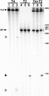
SegF prefers T2 DNA in vitro. Substrate DNAs that were end-labeled on their template (bottom) strands were generated by amplification of T4 DNA (primers 37 and 39) and of T2 DNA (primers 42 and 43). The positions of primers are shown in Figure 2C. Substrates were incubated singly (lanes 16) and together (lanes 79), with addition of in vitro synthesized SegF (lanes 3,6,9), a mock transcription/translation extract (lanes 2,5,8), or reaction buffer only (lanes 1,4,7). Cleavage products were resolved on a denaturing 6% polyacrylamide gel. Arrows indicate DNA substrates with the sizes indicated. Open triangles indicate the major cleavage products from a substrate with the T4 sequence; a solid triangle indicates the product from a substrate with the T2 sequence. Positions of the ΦX174/HaeIII DNA size standard are indicated on the right.
Figure 5
Figure 5
SegF preferentially cleaves T2 DNA in vivo. (A) E. coli B40 strr (Sup0) or B40 supF strr (Sup+) cells were infected with phage T2 in combination with phage T4 or T4am56Δ69 or T4am56am69. Total DNA was prepared from the mixture of phages before addition to cells (lanes 1,5,9) and from phage-infected cells 10 min (lanes 2,6,10,13), 20 min (lanes 3,7,11,14), and 30 min (lanes 4,8,12,15) after infection. The DNA was digested with restriction endonuclease PacI, separated by electrophoresis, and analyzed by Southern hybridization. The probe was a mixture of random-primer-labeled DNA that was generated by amplification of T2 and T4 DNAs with primers 37 and 26 (see Fig. 2C). Arrows indicate PacI restriction fragments containing the T2 and T4 target DNAs, and solid triangles indicate putative cleavage products from T2. The sizes shown to the left were approximated using a DNA ladder mix as size standards. Slower-migrating DNA species are indicated by X. (B) Blot A was stripped and reprobed with end-labeled T4-specific oligonucleotide 35 (see Fig. 2C). (C) Hybridization signals from the T4 target DNA (3.1-kb PacI fragment in wild type and T4am56am69 or 2.6-kb fragment from T4am56Δ69) and T2 target DNA (2.8-kb PacI fragment) from blot A were determined using a phosphorimager. The ratios of intensities of the T4 to T2 bands were determined and normalized with respect to the input ratio.
Figure 6
Figure 6
Nucleotide sequence alignment of gene 56 from T4 and T2. Nucleotide sequence alignment of gene 56 from phages T4 (accession no. AF158101) and T2 (Gary et al. 1998), based on the amino acid sequence alignment. Nucleotide gaps are indicated by dashes. Identical nucleotides between T4 and T2 are shaded. SegF cleavage on the coding strand of T2 gene 56 is indicated by an upward-pointing arrow. An asterisk indicates the position of a G → A change in T4am56E51 (Gary et al. 1998). A right-pointing arrow indicates the start of gene 69 in the +1 frame of the T4 gene 56 sequence.
Figure 7
Figure 7
Intron versus intronless homing. Filled boxes represent regions of DNA nonidentity. (A) Group I intron homing: An endonuclease encoded within the intron cleaves (↑) the intronless homolog close to the intron insertion site. DSB repair, which includes limited exonuclease digestion of cut DNA, incorporates the intron into the recipient, with frequency of coconversion of flanking markers (*) inversely proportional to their distance from the cleavage site. Insertion of the intron destroys the endonuclease recognition site. (B) Intronless homing by segF. SegF preferentially cleaves T2 gene 56, within a region of partial conservation with the T4 homolog (↑). Because of patchy conservation within gene 56, DSB repair initiates recombination in dam and soc, the closest regions of extensive sequence conservation. segF and the entire T4 gene 56 (with its nonpreferred SegF cleavage site) are transferred to recipients. The coconversion of markers in gene 56 (*) is close to 100%.

Similar articles

Cited by

References

    1. Altschul SF, Madden TL, Schaffer AA, Zhang J, Zhang Z, Miller W, Lipman DJ. Gapped BLAST and PSI-BLAST: A new generation of protein database search programs. Nucleic Acids Res. 1997;25:3389–3402. - PMC - PubMed
    1. Belfort M, Roberts RJ. Homing endonucleases: Keeping the house in order. Nucleic Acids Res. 1997;25:3379–3388. - PMC - PubMed
    1. Bell-Pedersen D, Quirk SM, Aubrey M, Belfort M. A site-specific endonuclease and co-conversion of flanking exons associated with the mobile td intron of phage T4. Gene. 1989;82:119–126. - PubMed
    1. Bell-Pedersen D, Quirk S, Clyman J, Belfort M. Intron mobility in phage T4 is dependent upon a distinctive class of endonucleases and independent of DNA sequences encoding the intron core: Mechanistic and evolutionary implications. Nucleic Acids Res. 1990;18:3763–3770. - PMC - PubMed
    1. Chevalier BS, Stoddard BL. Homing endonucleases: Structural and functional insight into the catalysts of intron/intein mobility. Nucleic Acids Res. 2001;29:3757–3774. - PMC - PubMed

Publication types

MeSH terms