. 2002 Feb 1;22(3):698-707.
doi: 10.1523/JNEUROSCI.22-03-00698.2002.

Process outgrowth of oligodendrocytes is promoted by interaction of fyn kinase with the cytoskeletal protein tau

Affiliations

Process outgrowth of oligodendrocytes is promoted by interaction of fyn kinase with the cytoskeletal protein tau

Corinna Klein et al. J Neurosci. .

Abstract

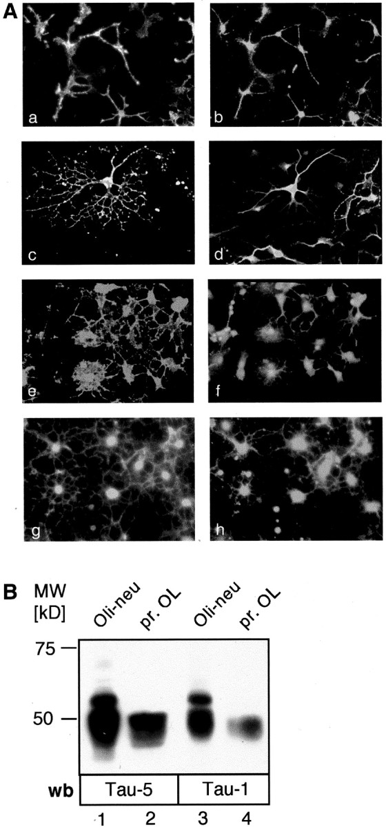
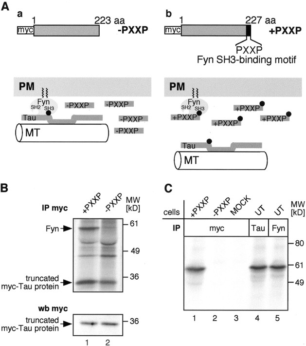
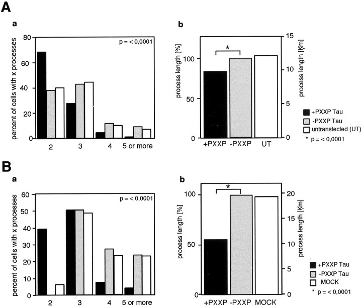
Fyn kinase plays an important role during myelination and has been shown to promote morphological differentiation of cultured oligodendrocytes. We analyzed the downstream targets of Fyn kinase in oligodendrocytes. Because process outgrowth and wrapping of axons involve cytoskeletal rearrangement, we focused on cytoskeletal proteins linked to Fyn. Here we demonstrate that Fyn binds to the cytoskeletal proteins Tau and alpha-Tubulin in oligodendrocytes. Tau interacts with the Fyn SH3 domain whereas alpha-Tubulin binds to the Fyn SH2 and SH3 domains. To study the function of the Fyn-Tau interaction in oligodendrocytes, we designed a Tau deletion mutant that would compete with endogenous Tau-Fyn binding in transfected cells. The mutant Tau protein binds to the Fyn SH3 domain but lacks the microtubuli interaction domain and thus cannot bind to microtubuli. In the presence of the mutant Tau protein, a reduction of the process number and process length in oligodendroglial cells was observed. This effect is likely to be caused by interference with the Fyn-Tau-microtubuli cascade rather than inactivation of the kinase, because Fyn bound to the mutant Tau retains activity. A similar inhibition of process outgrowth was observed when oliogodendroglial cells were cultured in the presence of Fumonisin B1, an inhibitor of sphingolipid synthesis that prevents the formation of rafts. Because ligation of the cell adhesion molecule F3 on oligodendrocytes leads to activation of Fyn kinase localized in rafts, these findings suggest that recruitment of Tau and Tubulin to activated Fyn kinase in rafts is an important step in the initiation of myelination.


Figures

Fig. 1.
Fig. 1.
Cultured oligodendrocytes express Tau.A, Immunofluorescence staining. Primary oligodendrocytes were stained with antibodies to oligodendroglial markers and Tau (af) or with Fyn and Tau (g, h). All AN2-positive oligodendrocyte precursor cells (a) stain for Tau (b). Tau is expressed in the cell soma as well as in the processes. Primary oligodendrocytes were costained with antibodies to the oligodendroglial markers O4 (c) or MAG (e) and Tau (d, f). Cell somata as well as large membrane processes are positive for Tau, whereas the tips of very small processes are unstained. O4 and MAG staining are distributed all over the cell surface including small processes and membrane protrusions. B, Western blot analysis. Both primary oligodendrocytes (pr. OL) and the precursor cell line Oli-neu express Tau. Cell lysates of Oli-neu cells (lanes 1, 3) and primary oligodendrocytes (lanes 2, 4) were resolved by SDS-PAGE and immunoblotted with the antibodies Tau-5 (lanes 1, 2) recognizing an unphosphorylated epitope and Tau-1 (lanes 3, 4) recognizing a dephosphorylated serine at aa 199 (human nomenclature). Several isoforms are detected by both antibodies running between 45 and 65 kDa. Oli-neu cells may express additional isoforms compared with primary oligodendrocytes, or alternatively the isoforms are subjected to different posttranslational modifications. wb, Western blot.
Fig. 2.
Fig. 2.
Fyn is associated with Tau and Tubulin in primary oligodendrocytes. Raft fractions and bottom gradient fractions of sucrose density gradients from primary oligodendrocytes were subjected to immunoprecipitation (IP) using antibodies to Fyn followed by SDS-PAGE (lanes 1, 2) or were analyzed directly by SDS-PAGE (lanes 3,4) followed by Western blot (wb) analysis using monoclonal antibodies against Fyn, Lyn, Tau, and α-Tubulin. Fyn associates with Tau and α-Tubulin in the raft fraction as well as in the bottom gradient fraction. Lyn, which is also found in the raft fraction (Lyn, lane 3), is not associated with this complex, either in the raft fraction (lane 1) or in the non-raft fraction (lane 2).
Fig. 3.
Fig. 3.
The SH3 and SH2 domains of Fyn interact with cytoskeletal proteins. Primary oligodendrocyte lysates were incubated with the different GST-fusion proteins and subjected to SDS-PAGE followed by Western blot analysis (wb). Tau precipitates specifically with fusion proteins containing the SH3 domain of Fyn (lane 4); α-Tubulin precipitates with the fusion proteins containing the SH2 or SH3 domain of Fyn (lanes 3, 4). Neither Tau nor α-Tubulin binds to the SH3 domain of Amphiphysin (lane 2) or to GST alone (lane 1).
Fig. 4.
Fig. 4.
Tau deletion protein +PXXP bearing the Fyn binding motif binds to active Fyn. A, Design of the two rat Tau deletion mutants and model of action. a, −PXXP (aa 1–223), lacking the Fyn SH3 binding motif on Tau;b, +PXXP (aa 1–227), including the Fyn SH3 binding motif (PXXP). Both constructs carry a myc tag at the N terminus. When overexpressed in oligodendroglial cells, the +PXXP Tau protein competes with endogenous Tau for Fyn binding. Because the +PXXP Tau protein is lacking the microtubuli (MT) binding domain, the Fyn–Tau–Tubulin cascade is disrupted (b). In contrast, when the −PXXP control protein is overexpressed in these cells, the interaction cascade is preserved (a). B, Immunoprecipitation of radiolabeled Oli-neu cells with antibodies to myc. Oli-neu cells expressing either the +PXXP or −PXXP Tau protein were metabolically labeled with 35S-methionine/cysteine and subjected to immunoprecipitation (IP) with antibodies against myc followed by SDS-PAGE and Phosphoimager analysis. From cells expressing the +PXXP construct, a signal for Fyn at 59 kDa is seen (top panel, arrow, lane 1). From cells expressing the −PXXP construct, no signal is seen (lane 2). Equal numbers of transfected cells were loaded on SDS-PAGE and analyzed by Western blot (wb) with antibodies against myc (bottom panel), demonstrating a similar expression level of each Tau mutant.C, Kinase assays on the myc immunoprecipitates. Oli-neu cells were transiently transfected with +PXXP (lane 1), −PXXP (lane 2), or empty vector (MOCK;lane 3) and were subjected to immunoprecipitation with antibodies to myc. As a control, untransfected Oli-neu cells were subjected to immunoprecipitation with antibodies to Tau (lane 4) and Fyn (lane 5). An in vitro kinase assay was performed on the precipitates followed by SDS-PAGE and autoradiography. A strong signal for phosphorylated and thus active Fyn is present in the myc precipitates from cells expressing the +PXXP construct (lane 1) and in the Tau immunoprecipitates (lane 4).
Fig. 5.
Fig. 5.
Inhibition of the interaction between Fyn and Tau decreases process number and process length in oligodendroglial cells. Oli-neu cells (A) or primary oligodendrocytes (B) were transfected with the Tau deletion constructs −PXXP and +PXXP and cultured for 1 d. The process length and number of processes were analyzed. Processes longer than half the diameter of the cell soma were included in the measurements. Three individual experiments were performed, and the data were summarized and analyzed by the Mann–Whitney rank sum test. Oli-neu cells (A,a;p < 0.0001) as well as primary oligodendrocytes (B,a; p< 0.0001) expressing the +PXXP construct have fewer processes compared with the −PXXP transfected controls. The process length of +PXXP transfected Oli-neu cells is reduced by 16% (A,b; p< 0.0001), and the process length of +PXXP transfected primary oligodendrocytes is reduced by 49% (B,b; p< 0.0001) compared with −PXXP expressing cells and untransfected (UT) or MOCK transfected cells.
Fig. 6.
Fig. 6.
Inhibition of glycosphingolipid synthesis prevents raft formation in oligodendrocytes and inhibits process growth in Oli-neu cells. A, Distribution of NCAM protein and lipids on gradients of FB1-treated oligodendrocytes. a, Primary oligodendrocytes were cultured for 5 d with Fumonisin B1 (FB1) and fractionated on sucrose density gradients. The fractions were collected and analyzed by Western blot with antibodies against NCAM (a) or were subjected to lipid extraction and analyzed by TLC (b). Fraction (Frakt.) 1 is the bottom gradient fraction. In cells cultured in the presence of FB1, the GPI-anchored NCAM form NCAM 120 is no longer localized in the raft fraction as is seen in the untreated cells (a), but is entirely present in the non-raft fraction. b, FB1-treated primary oligodendrocytes do not synthesize sphingolipids. No signal for galactocerebroside (GalC) or Sulfatide (Sulf) can be detected in lipids of FB1-treated cells in comparison with control cells. Chol, Cholesterol; PE, phosphatidylethanolamine;SM, sphingomyelin. B, FB1 inhibits process outgrowth of Oli-neu cells. a, Oli-neu cells were cultured for 3 d with FB1 (+) or without FB1 (−). Raft fractions and non-raft fractions were collected from sucrose density gradients and subjected to SDS-PAGE followed by Western blot analysis with antibodies against Tau. Culture in FB1 results in the absence of Tau from low density gradient fractions; in untreated cells Tau is localized in rafts in these gradient fractions. Oli-neu cells were cultured in the absence of (b) or in the presence of (c) 50 μm FB1 for several days (see Material and Methods). After 5 d in culture in FB1 including one passage, the cells are almost lacking processes (c). Nevertheless they are still alive, because after further culture (2 d) in the absence of FB1 the cells recover and regenerate processes (d).
Fig. 7.
Fig. 7.
Proposed model of the role of Fyn and Tau in myelination. During development, oligodendrocyte processes initiate contact with an axon. The interaction of adhesion molecules such as F3 and signaling events in both cells are necessary (1.). Ligation of F3 by an axonal ligand activates Fyn in raft microdomains leading to increased binding of Tau. Microtubuli (MT) are recruited to the area of contact (2.). Vesicular transport to the contact site is facilitated (3.), and insertion of vesicles containing myelin-specific lipid and proteins promotes further internodal process elongation along and around the axon (4.).PM, Plasma membrane; GSL, glycosphingolipid; Chol, cholesterol.

Similar articles

Cited by

References

    1. Biffiger K, Bartsch S, Montag D, Aguzzi A, Schachner M, Bartsch U. Severe hypomyelination of the murine CNS in the absence of myelin-associated glycoprotein and fyn tyrosine kinase. J Neurosci. 2000;20:7430–7437. - PMC - PubMed
    1. Binder LI, Frankfurter A, Rebhun LI. The distribution of tau in the mammalian central nervous system. J Cell Biol. 1985;101:1371–1378. - PMC - PubMed
    1. Bligh EG, Dyer WJ. A rapid method of total lipid extraction and purification. Can J Biochem Physiol. 1957;37:911–917. - PubMed
    1. Brandt R. The tau proteins in neuronal growth and development. Front Biosci. 1996;1:d118–130. - PubMed
    1. Brandt R, Leger J, Lee G. Interaction of tau with the neural plasma membrane mediated by tau's amino-terminal projection domain. J Cell Biol. 1995;131:1327–1340. - PMC - PubMed

Publication types

MeSH terms