. 2002 Feb 1;22(3):748-56.
doi: 10.1523/JNEUROSCI.22-03-00748.2002.

Ultrastructure of a somatic spine mat for nicotinic signaling in neurons

Affiliations

Ultrastructure of a somatic spine mat for nicotinic signaling in neurons

Richard D Shoop et al. J Neurosci. .

Abstract

Chick ciliary neurons have somatic spines grouped in discrete clumps or mats tightly folded against the soma and enriched in nicotinic receptors containing alpha7 subunits. An embryonic ciliary neuron has one to two dozen such spine mats, all overlaid by a large presynaptic calyx engulfing the cell. Three-dimensional tomographic reconstruction from serial thick sections revealed 13 somatic spines in one complete spine mat on a ciliary neuron late in embryogenesis. The spines varied in morphology and usually were branched but had numerous similarities to dendritic spines, including mean length, volume, surface area, presence of endoplasmic reticulum, and occasional multivesicular bodies. The spines invariably were connected to the soma via a narrow neck of approximately 0.2 micrometer in diameter as found for dendritic spines, suggesting restricted access from spine lumen to soma. A prominent difference between dendritic and somatic spines is the absence of postsynaptic densities from most somatic spines both on embryonic and adult ciliary neurons. Transmitter access to receptors on the spines may occur either by lateral diffusion from release sites over nearby postsynaptic densities or by release directly onto spines from the overlying calyx lined with vesicles. The latter is less likely in the adult, where some spines are adjacent to but not overlaid by vesicle-enriched presynaptic structures. The anatomical configuration of spine mats suggests coordinate spine activation by transmitter release into a confined volume while spine morphology is used to control the chemical consequences of synaptic signaling.


Figures

Fig. 1.
Fig. 1.
Traditional EM on thin sections of E15 ciliary neurons showing spines containing PSDs and ER and being surrounded by vesicle-filled presynaptic calyx. A, Spine mat showing spines (sp) with PSDs (arrowheads) and ER (small arrow) lying between the neuron soma (n) and presynaptic calyx (c). A calyx protrusion extends into the mat but is distinct from the two spines, although the intervening membranes are not always readily visible in isolated thin sections. B, A spine mat lying between a neuron soma (n) and overlying calyx (c) and showing a spine neck connecting a spine to the soma (large arrow) plus a spine (sp) with ER (small arrow). Scale bar, 500 nm.
Fig. 2.
Fig. 2.
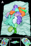
A complete spine mat with 13 somatic spines on an E15 ciliary neuron. A, The spine mat is shown at a 45° angle from the horizontal, with spines individually color-coded (in this and subsequent figures) against the soma membrane shown ingray and covering a 24 μm2 area.B, Rotating the spine mat by 200° shows the opposite side of the spine mat. C, Tilting the neuronal surface to examine the membrane on edge shows that the somatic spines lie in a cavity in the neuronal soma and also project above the soma surface.D, Rotating the view in C by 90° in the horizontal gives a view from directly above the spine mat. Scale bars, 500 nm.
Fig. 3.
Fig. 3.
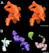
Selected individual spines illustrating the diverse morphologies. A, One of the largest and most branched somatic spines of the E15 mat, shown as a stereo pair.B, Four additional examples of somatic spines from the same mat, each shown at three-fourths the size of that inA. Spine necks and attachment sites are indicated ingreen. Scale bars, 500 nm.
Fig. 4.
Fig. 4.
Characteristic attachment sites and contents of somatic spines on an E15 ciliary neuron. A, Soma membrane (gray; as in Fig.2A) with the spines removed and the attachment sites indicated in green. The attachment sites are approximately the same size and are distributed nonuniformly over the entire surface area supporting the spine mat. B, C, Examples from reconstructed cross sections showing spine necks and attachment sites (arrowheads) to the soma. Multivesicular bodies (asterisk) were seen sometimes.D, E, Examples from reconstructed cross sections showing ER (arrowheads), which was extensive in seven of the 13 spines. F, Semitransparent rendition of spine 11 to illustrate the internal distribution of ER (gray). Scale bars: A, 500 nm;B–F, 200 nm.
Fig. 5.
Fig. 5.
Distribution of PSDs on and around an E15 spine mat. A, Only four of the 13 spines had a PSD (yellow), although a fifth PSD was located on the soma membrane nearby. B–D, All PSDs had the traditional structure, including a postsynaptic thickening (arrowheads) and closely apposed presynaptic membrane. None of the PSDs on somatic spines was associated with a distinct spine head. Scale bars: A, 500 nm; D, 200 nm.
Fig. 6.
Fig. 6.
Distribution of synaptic vesicles in the presynaptic calyx overlaying the E15 spine mat. A, All synaptic vesicles in the serial sections containing the spine mat were marked individually (red) and added to the reconstructed spine mat image from Figure 2A. B, Side view indicating the vertical distribution of the vesicle population. C, Side view with the postsynaptic membrane removed, illustrating vesicle-containing structures (arrowhead) extending into the spine mat.D, Deleting all synaptic vesicles ≥5 nm from the presynaptic membrane to reveal potentially docked vesicles at release sites left significant concentrations over the PSDs (yellow), as expected. In addition, scattered vesicles remained over much of the spine surface area, suggesting possible docked vesicles even in the absence of juxtaposed PSDs. Scale bar, 500 nm. The artificial diagonal border of vesicles in thetop rightcorners of B andC is attributable to the boundary of the visualized volume, not the perimeter of the calyx.
Fig. 7.
Fig. 7.
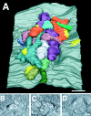
A nearly complete spine mat reconstructed from an adult ciliary neuron. A, The spine mat is shown with spines individually color-coded (in this and subsequent figures) against the soma membrane (gray) covering a 24 μm2 area. B, The attachment sites of the spine necks are shown in green after the spines were removed from the reconstructed area shown in A. A block-like outpocketing of the soma membrane, clearly different from the spines, is also present; only one such outpocketing was found among the several tomograms that were examined. C, Individually reconstructed spines have morphologies as diverse as those at E15. Scale bars: A, B, 500 nm; C, 200 nm.
Fig. 8.
Fig. 8.
Distribution of synaptic vesicles in the presynaptic calyx overlaying an adult spine mat. A, All synaptic vesicles in the serial sections encompassing the spine mat were marked individually (red) and are shown superimposed on the spine mat from Figure 7. None of the spines has PSDs; unlike the situation at the embryonic spine mat, very few vesicles overlay the individual adult spines. B, The same view as in A showing the synaptic vesicles after the spines have been removed. The large pool of synaptic vesicles in the lower right quadrant lies adjacent to the spine mat. Scale bars, 500 nm.
Fig. 9.
Fig. 9.
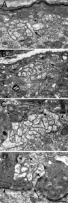
EM thin sections of adult ciliary neurons showing spines mats. A, B, Spine mats lying between a neuron (n) and the presynaptic calyx (c). Although the spines (sp) have ER (small arrows), in these examples they do not have PSDs and are not adjacent to presynaptic structures with appreciable numbers of vesicles. A spine neck is indicated by the large arrow in A. C, D, Spine mats showing both ER (small arrow) and synaptic vesicles in the overlying calyx (c). Multiple PSDs are indicated (arrowheads). The spine mat in D is surrounded totally by a large pool of vesicles, in sharp contrast to the almost complete lack of vesicles in most thin sections through the spine mats in A andB. Scale bar, 500 nm.
None

References

    1. Alkondon M, Albuquerque EX. Diversity of nicotinic acetylcholine receptors in rat hippocampal neurons. I. Pharmacological and functional evidence for distinct structural subtypes. J Pharmacol Exp Ther. 1993;265:1455–1473. - PubMed
    1. Alkondon M, Pereira EFR, Cortes WS, Maelicke A, Albuquerque EX. Choline is a selective agonist of α7 nicotinic acetylcholine receptors in rat brain neurons. Eur J Neurosci. 1997;9:2734–2742. - PubMed
    1. Bundman MC, Pico RM, Gall CM. Ultrastructural plasticity of the dentate gyrus granule cells following recurrent limbic seizures. I. Increase in somatic spines. Hippocampus. 1994;4:601–610. - PubMed
    1. Chang K, Berg DK. Dependence of circuit function on nicotinic acetylcholine receptors containing α7 subunits. J Neurosci. 1999;19:3701–3710. - PMC - PubMed
    1. Chang K, Berg DK. Voltage-gated channels block nicotinic regulation of CREB phosphorylation and gene expression in neurons. Neuron. 2001;32:855–865. - PubMed

Publication types

MeSH terms

Substances

LinkOut - more resources