. 2002 Feb 1;22(3):842-53.
doi: 10.1523/JNEUROSCI.22-03-00842.2002.

Repellent guidance of regenerating optic axons by chondroitin sulfate glycosaminoglycans in zebrafish

Affiliations

Repellent guidance of regenerating optic axons by chondroitin sulfate glycosaminoglycans in zebrafish

Catherina G Becker et al. J Neurosci. .

Abstract

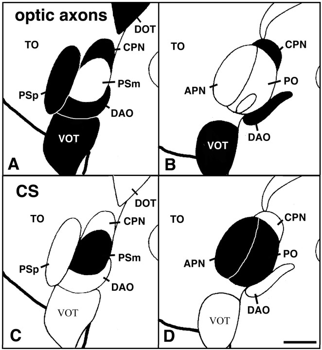
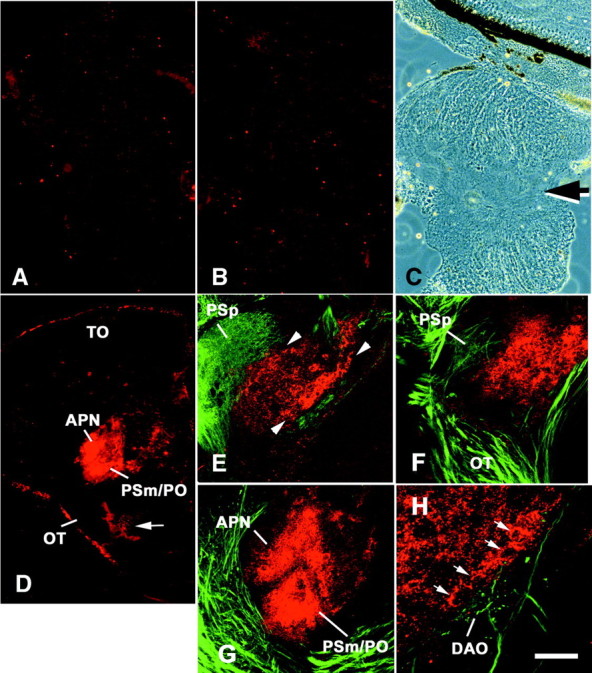
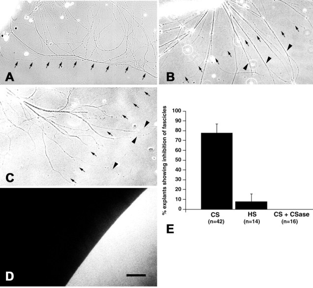
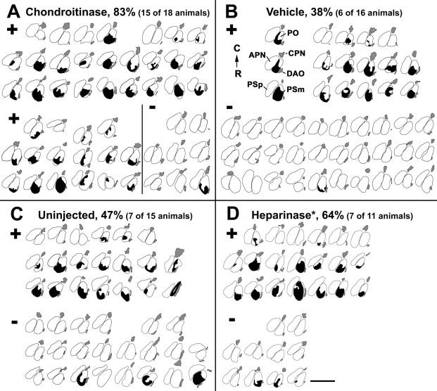
We analyzed the role of chondroitin sulfate (CS) glycosaminoglycans, putative inhibitors of axonal regeneration in mammals, in the regenerating visual pathway of adult zebrafish. In the adult, CS immunoreactivity was not detectable before or after an optic nerve crush in the optic nerve and tract but was constitutively present in developing and adult nonretinorecipient pretectal brain nuclei, where CSs may form a boundary preventing regenerating optic fibers from growing into these inappropriate locations. Enzymatic removal of CSs by chondroitinase ABC after optic nerve crush significantly increased the number of animals showing erroneous growth of optic axons into the nonretinorecipient magnocellular superficial/posterior pretectal nucleus (83% vs 42% in controls). In vitro, a substrate border of CSs, but not heparan sulfates, strongly repelled regenerating retinal axons from adult zebrafish. We conclude that CSs contribute to repellent axon guidance during regeneration of the optic projection in zebrafish.


Figures

Fig. 1.
Fig. 1.
Semischematic representation of the distribution of optic fibers (A, B) and CS (C, D) in the pretectum of adult zebrafish. The same two consecutive transverse sections are shown in A and B andC and D; dorsal is at thetop; lateral is left. Aand C are 60 μm rostral to B andD. The presence of optic fibers in A andB and CS immunoreactivity in C andD is indicated by black filling of brain structures. Optic fibers are present in the parvocellular superficial pretectal nucleus (PSp), the central pretectal nucleus (CPN), the dorsal accessory optic nucleus (DAO); the optic tectum (TO; innervation not indicated), and the ventral optic tract (VOT) and dorsal optic tract (DOT). The magnocellular superficial pretectal nucleus (PSm), the accessory pretectal nucleus (APN), and the posterior pretectal nucleus (PO) are free of optic fibers but are strongly CS-immunopositive. Outlines of brain nuclei are taken fromWullimann et al. (1996). Scale bar, 100 μm.
Fig. 2.
Fig. 2.
CS immunoreactivity is not increased after optic nerve crush but is constitutively present in specific pretectal brain nuclei. A–C, Longitudinal sections through the optic nerve are shown; the retina is at the top. CS immunoreactivity is low in the unlesioned optic nerve (A) and is not altered 7 d after an optic nerve crush (B). The crush site is indicated by an arrow in the phase-contrast image inC, corresponding to B. D, Cross section through a brain. Dorsal is at the top; lateral is left. CS immunoreactivity in an unlesioned animal is very low in the optic tract (OT) and the tectum (TO) but intense in the magnocellular superficial/posterior pretectal nucleus (PSm/PO) and the accessory pretectal nucleus (APN). Thearrow points to CS-immunopositive meninges.E–H, Visualization of biocytin-labeled optic fibers (green) and CS immunoreactivity (red) using confocal laser scanning microscopy. Orientations are the same as in D.E, The rostral magnocellular superficial pretectal nucleus (red) is contiguous with the parvocellular superficial pretectal nucleus (PSp), which receives dense retinal fibers in an unlesioned animal. CS immunoreactivity is more intense at the border of the magnocellular superficial pretectal nucleus (arrowheads) than in its center.F, Three weeks after a lesion, CS immunoreactivity in the rostral (Figure legend continued.) magnocellular superficial pretectal nucleus is comparable with that in unlesioned controls, and optic fibers have grown back through the optic tract (OT) and reinnervate the parvocellular superficial pretectal nucleus (PSp). G, In a more caudal cross section through the diencephalon, the accessory pretectal nucleus (APN) and the magnocellular superficial/posterior pretectal nucleus (PSm/PO) are strongly labeled by CS antibodies 3 weeks after the lesion. Regenerating fibers grow around these nuclei. H, At a higher magnification, intensely CS-immunopositive cells (arrows) are detectable at the medial border of the magnocellular superficial/posterior pretectal nucleus in an animal 3 weeks after an optic nerve crush. Fibers with small protrusions, which are probably terminals in the dorsal accessory optic nucleus (DAO) and smooth fibers, which are probably fibers of passage, grow along this boundary. Scale bar, 100 μm (forA–C), 200 μm (for D), 75 μm (forE–G), 25 μm (for H).
Fig. 3.
Fig. 3.
CS immunoreactivity is present in the pretectum during development. Cross sections through whole larvae are shown; dorsal is at the top; arrowheads inA and B indicate the brain midline; inC and D, lateral is left.A, C, At 8 d of development, weak CS immunoreactivity is present in the pretectum (arrows). At higher magnification (C), the characteristic small punctate appearance of CS labeling is visible (C, arrow). Large spots of immunoreactivity (C, arrowheads) are an artifact from material that separated from the intensely immunopositive cartilage (A, asterisks). Meninges are also CS-immunopositive (C, asterisks).B, D, By 28 d of development, immunoreactivity is distributed in a ring-like pattern in the lateral diencephalon (arrows), resembling the adult configuration.D is a higher magnification of B. Cartilage (D, asterisk) is intensely labeled. Scale bars: A, B, 100 μm; C, D, 50 μm.
Fig. 4.
Fig. 4.
A substrate border of CSs but not HSs repels regenerating optic axons in vitro. A–D, Substrate borders of CSs (A), HSs (B), and CSs after chondroitinase treatment (C) are indicated by small arrows. The position of the substrate border was visualized under fluorescence optics as shown in D, which is taken from the same area depicted in C. Fibers grow from retinal explants that are located in the top left corner. Although fibers are deflected at a CS border (A), they readily invade a substrate spot of HSs (B). The repellent activity of a CS border is abolished after treatment of the substrate with chondroitinase (C).Arrowheads in B and Cindicate fibers that crossed the substrate border. E, Quantification of the percentage of explants showing deflection of axons at a substrate border. Inhibition of axon growth at a CS border was statistically highly significant compared with HS borders or chondroitinase (CSase)-digested CS substrate spots (Fisher's exact test, p ≤ 0.0003;n = number of explants observed). Scale bar, 100 μm (for A–D).
Fig. 5.
Fig. 5.
CS immunoreactivity but not tenascin-R immunoreactivity is removed from the magnocellular superficial pretectal nucleus of adult zebrafish in vivo by different enzymes. Cross sections are shown; dorsal is at thetop; lateral is left.A–D, At 1 d after chondroitinase injection (B), no CS immunoreactivity is detectable with antibody CS-56 in the magnocellular superficial pretectal nucleus compared with uninjected controls (A).C, Phase-contrast image corresponding toB. D, At 7 d after injection, weak CS immunoreactivity is detectable with antibody CS-56 around large neurons in the magnocellular superficial pretectal nucleus (arrows). However, immunoreactivity is generally considerably lower than in uninjected controls (A). E, F, At 1 d after chondroitinase injection (F), chondroitin sulfate stub immunoreactivity, indicating successful removal of CSs, is increased in the magnocellular pretectal nucleus compared with uninjected controls (E). G, H, Chondroitinase injection does not alter tenascin-R immunoreactivity 1 d after injection (H) compared with uninjected controls (G). I, J, CS immunoreactivity is reduced but still detectable in the magnocellular superficial pretectal nucleus 1 d after heparinase injection (J) compared with uninjected controls that were processed on the same slide (I). Scale bar, 100 μm.
Fig. 6.
Fig. 6.
Retinal ganglion cell axons invade the magnocellular superficial/posterior pretectal nucleus after chondroitinase treatment. Vibratome cross sections (50 μm in thickness) through the brain are shown. Optic fibers are labeled with biocytin in brown. Cell somata are counterstained with neutral red; dorsal is at the top; lateral isleft. No fibers are detectable in the magnocellular superficial/posterior pretectal nucleus (PSm/PO) 3 weeks after a lesion of the contralateral optic nerve in a vehicle injected animal at low (A) and high magnification (B). In a chondroitinase-treated animal, fibers are present in the magnocellular superficial/posterior pretectal nucleus 3 weeks after a lesion of the contralateral optic nerve, depicted at low (C) and high magnification (D) of the same section. In the vehicle- and chondroitinase-injected cases, fibers are present in the central pretectal nucleus (A, C, CPN) and the dorsal accessory optic nucleus (B, D, DAO), which served as an internal control for efficient labeling of the optic projection. Note that the section in C includes a part of the parvocellular superficial pretectal nucleus (PSp), which is reinnervated by optic fibers, whereas the section depicted inA is slightly more caudal and contains the accessory pretectal nucleus (APN) next to the magnocellular superficial/posterior pretectal nucleus (PSm/PO). Scale bars: C, 100 μm (for A, C); D, 40 μm (for B, D).
Fig. 7.
Fig. 7.
Outlines of the areas invaded by regenerating optic fibers in sections of the magnocellular superficial/posterior pretectal nucleus after different treatments. Chartings of all cases that received chondroitinase (A), vehicle (B), no injection (C), or heparinase injection (D) after contralateral optic nerve crush are shown; dorsal is at the top; lateral isleft. The magnocellular superficial/posterior pretectal nucleus stretches over two to three cross sections. These are depicted in columns for the individual cases. All chartings are organized as in the first case in B, with the most rostral section on the bottom and the most caudal section on the top (RC). The parvocellular superficial (PSp), magnocellular superficial (PSm), accessory (APN), central (CPN), and posterior (PO) pretectal nuclei, as well as the dorsal accessory optic nucleus (DAO), are outlined as indicated for the first case in B. The area taken by fibers invading the magnocellular superficial/posterior pretectal nucleus in cross sections is black. Fibers reinnervating their regular terminal fields in the dorsal accessory optic nucleus and the central pretectal nucleus are gray. Fibers reinnervating the parvocellular superficial pretectal nucleus after a lesion have been omitted for clarity. Animals were scored as having fibers invading the magnocellular superficial/posterior pretectal nucleus when fibers were present in these nuclei in at least two consecutive sections (see Materials and Methods). Cases are sorted accordingly (+, invasion of fibers; −, no invasion of fibers), and the percentages of cases with fibers in the magnocellular superficial/posterior pretectal nucleus are given. The proportion of cases with fibers in the magnocellular superficial/posterior pretectal nucleus after chondroitinase treatment is highly significantly increased (p = 0.003) compared with vehicle-injected and uninjected controls. *Note that the heparinase preparation contained chondroitinase activity (see Results). Scale bar, 200 μm.

Similar articles

Cited by

References

    1. Anderson RB, Walz A, Holt CE, Key B. Chondroitin sulfates modulate axon guidance in embryonic Xenopus brain. Dev Biol. 1998;202:235–243. - PubMed
    1. Avnur Z, Geiger B. Immunocytochemical localization of native chondroitin-sulfate in tissues and cultured cells using specific monoclonal antibodies. Cell. 1984;38:811–822. - PubMed
    1. Bastmeyer M, Beckmann M, Schwab ME, Stuermer CAO. Growth of regenerating goldfish axons is inhibited by rat oligodendrocytes and CNS myelin but not by goldfish optic nerve tract oligodendrocyte-like cells and fish CNS myelin. J Neurosci. 1991;11:626–640. - PMC - PubMed
    1. Battisti WP, Shinar Y, Schwartz M, Levitt P, Murray M. Temporal and spatial patterns of expression of laminin, chondroitin sulphate proteoglycan and HNK-1 immunoreactivity during regeneration in the goldfish optic nerve. J Neurocytol. 1992;21:557–573. - PubMed
    1. Becker CG, Becker T, Meyer RL, Schachner M. Tenascin-R inhibits the growth of optic fibers in vitro but is rapidly eliminated during optic nerve regeneration in the salamander Pleurodeles waltl. J Neurosci. 1999;19:813–827. - PMC - PubMed

Publication types

MeSH terms

LinkOut - more resources