

Dissociable neural responses related to pain intensity, stimulus intensity, and stimulus awareness within the anterior cingulate cortex: a parametric single-trial laser functional magnetic resonance imaging study
- PMID: 11826125
- PMCID: PMC6758484
- DOI: 10.1523/JNEUROSCI.22-03-00970.2002
Dissociable neural responses related to pain intensity, stimulus intensity, and stimulus awareness within the anterior cingulate cortex: a parametric single-trial laser functional magnetic resonance imaging study
Abstract
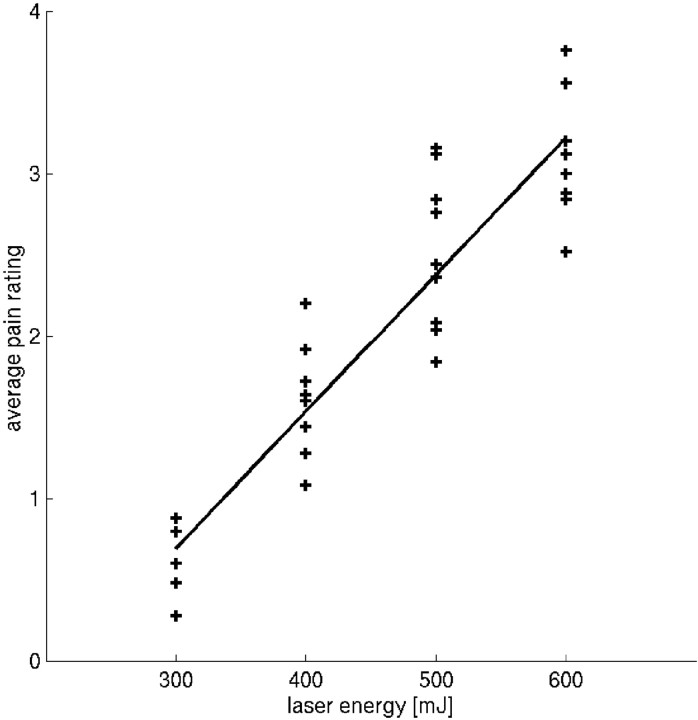
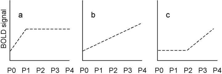
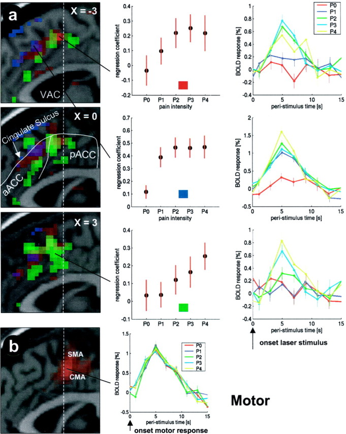
Neuroimaging studies have demonstrated activations in the anterior cingulate cortex (ACC) related to the affective component of pain, but not to stimulus intensity. However, it is possible that the low spatial resolution of positron emission tomography, as used in the majority of these studies, obscured areas coding stimulus intensity. We revisited this issue, using a parametric single-trial functional magnetic resonance imaging design, and investigated pain, stimulus intensity, and stimulus awareness (i.e., pain unrelated) responses within the ACC in nine healthy volunteers. Four different stimulus intensities ranging from warm to painful (300-600 mJ) were applied with a thulium yttrium-aluminum granite infrared laser in a randomized order and rated by the subjects on a five point scale (P0-P4). Pain-related regions in the ventral posterior ACC showed a response that did not distinguish between innocuous trials (P0 and P1) but showed a positive linear relationship with the blood oxygenation level-dependent contrast signal for painful trials (P2-P4). Regions in the dorsal anterior ACC along the cingulate sulcus differentiated between P0 (not perceived) and P1 but exhibited no additional signal increase with P2; these regions are related to stimulus awareness and probably to cognitive processing. Most importantly, we identified a region in the dorsal posterior ACC showing a response that discriminated between nonpainful trials (P0 and P1); therefore, this region was simply related to basic sensory processing and not to pain intensity. Stimulus-related activations were all located adjacent to the cingulate motor area, highlighting the strategic link of stimulus processing and response generation in the posterior ACC.
Figures
References
-
- Bouckoms AF. Psychosurgery for pain. In: Wall PD, Melzack R, editors. Textbook of pain. Churchill Livingstone; Edinburgh: 1989. pp. 868–881.
-
- Bromm B, Lorenz J. Neurophysiological evaluation of pain. Electroencephalogr Clin Neurophysiol. 1998;107:227–253. - PubMed
-
- Bromm B, Treede RD. Laser-evoked cerebral potentials in the assessment of cutaneous pain sensitivity in normal subjects and patients. Rev Neurol. 1991;147:625–643. - PubMed
-
- Büchel C, Dolan RJ. Classical fear conditioning in functional neuroimaging. Curr Opin Neurobiol. 2000;10:219–223. - PubMed
-
- Büchel C, Holmes AP, Rees G, Friston KJ. Characterizing stimulus-response functions using nonlinear regressors in parametric fMRI experiments. NeuroImage. 1998;8:140–148. - PubMed
Publication types
MeSH terms
LinkOut - more resources
Full Text Sources
Medical