

Distribution and behaviour of glabrous cutaneous receptors in the human foot sole
- PMID: 11826182
- PMCID: PMC2290100
- DOI: 10.1113/jphysiol.2001.013087
Distribution and behaviour of glabrous cutaneous receptors in the human foot sole
Abstract
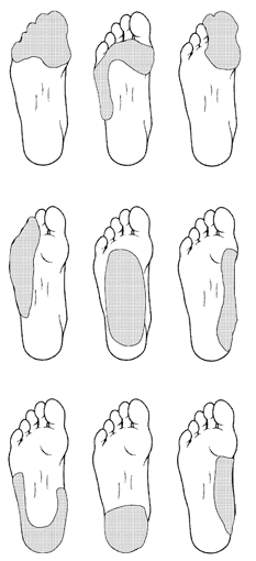
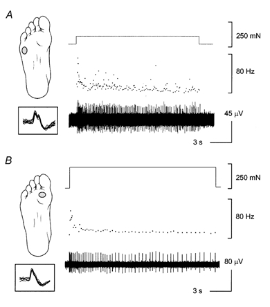
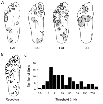
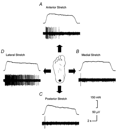
To document the activity of cutaneous mechanoreceptors in the glabrous skin of the foot sole, tungsten microelectrodes were inserted through the popliteal fossa and into the tibial nerve of thirteen healthy human subjects. A total of 104 cutaneous mechanoreceptors were identified in the glabrous skin of the foot. This sample consisted of 15 slow adapting type I (14 %), 16 slow adapting type II (15 %), 59 fast adapting type I (57 %), and 14 fast adapting type II units (14 %). The location of the receptors and the outline of the receptive fields were determined by using nylon monofilaments perpendicularly applied against the surface of the skin. This revealed that the receptors were widely distributed without an accumulation of receptors in the toes. There were also larger receptive fields predominantly isolated on the plantar surface of the metatarsal-tarsal region of the foot sole. Furthermore, with the foot in an unloaded position, there was no background discharge activity in any of the cutaneous receptors in the absence of intentionally applied stimulation. These findings suggest that skin receptors in the foot sole behave differently from those receptors found on the glabrous skin of the hand. This may reflect the role of foot sole skin receptors in standing balance and movement control.
Figures
References
-
- Allum JH, Bloem BR, Carpenter MG, Hulliger M, Hadders-Algra M. Proprioceptive control of posture: a review of new concepts. Gait and Posture. 1998;8:214–242. - PubMed
-
- Asai H, Fujiwara K, Toyama H, Yamashina T, Tachino K, Nara I. The influence of foot soles cooling on standing postural control analyzed by tracking the center of foot pressure. In: Woollacoot M, Horak F, editors. Posture and Gait: Control Mechanisms. II. Eugene, OR, USA: University of Oregon Books; 1992. pp. 151–154.
-
- Do MC, Bussel B, Breniere Y. Influence of plantar cutaneous afferents on early compensatory reactions to forward fall. Experimental Brain Research. 1990;79:319–324. - PubMed
-
- Edin BB. Quantitative analysis of static strain sensitivity in human mechanoreceptors from hairy skin. Journal of Neurophysiology. 1992;67:1105–1113. - PubMed
Publication types
MeSH terms
LinkOut - more resources
Full Text Sources
Other Literature Sources
Medical