. 2002 Jan;14(1):149-63.
doi: 10.1105/tpc.010301.

Arabidopsis CAP regulates the actin cytoskeleton necessary for plant cell elongation and division

Affiliations

Arabidopsis CAP regulates the actin cytoskeleton necessary for plant cell elongation and division

Roberto A Barrero et al. Plant Cell. 2002 Jan.

Abstract

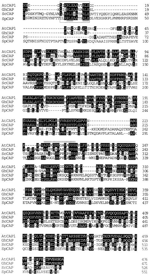
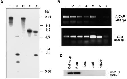
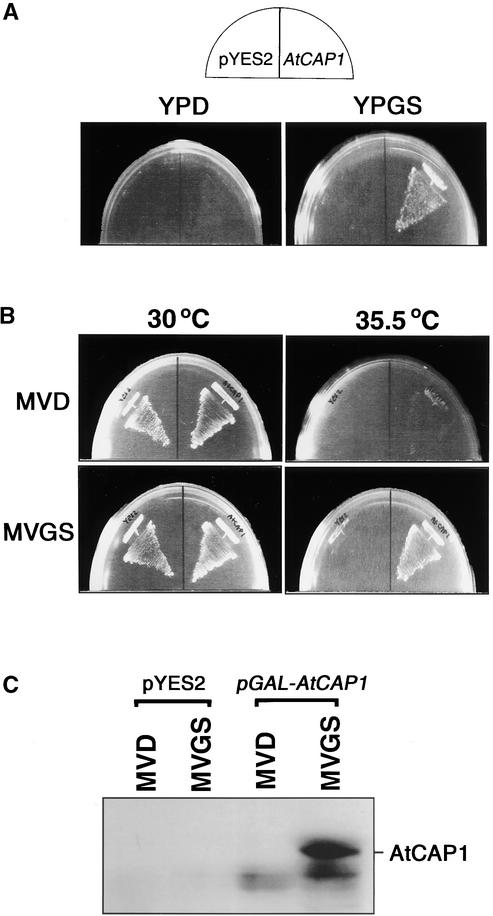
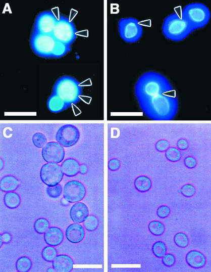
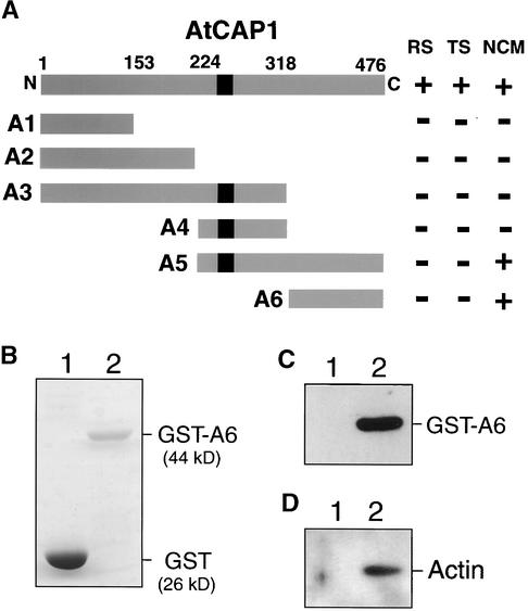
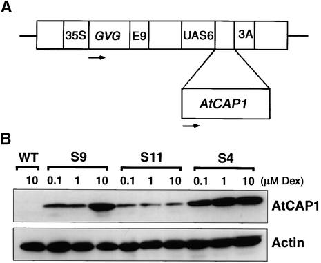
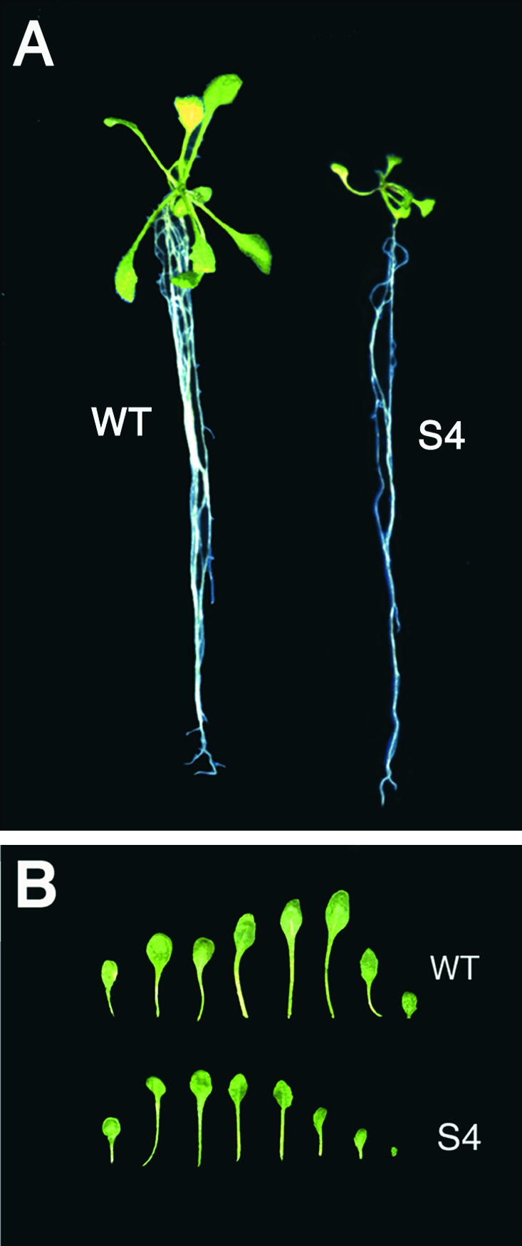
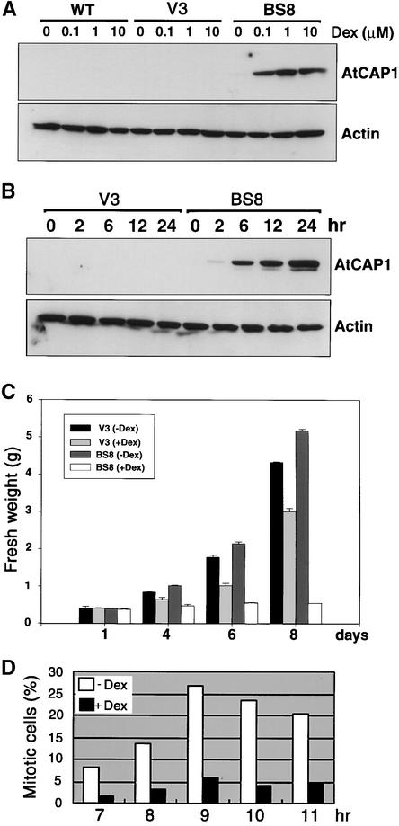
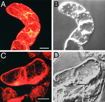
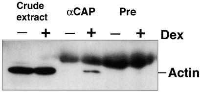
An Arabidopsis cDNA (AtCAP1) that encodes a predicted protein of 476 amino acids highly homologous with the yeast cyclase-associated protein (CAP) was isolated. Expression of AtCAP1 in the budding yeast CAP mutant was able to rescue defects such as abnormal cell morphology and random budding pattern. The C-terminal domain, 158 amino acids of AtCAP1 possessing in vitro actin binding activity, was needed for the regulation of cytoskeleton-related defects of yeast. Transgenic plants overexpressing AtCAP1 under the regulation of a glucocorticoid-inducible promoter showed different levels of AtCAP1 accumulation related to the extent of growth abnormalities, in particular size reduction of leaves as well as petioles. Morphological alterations in leaves were attributable to decreased cell size and cell number in both epidermal and mesophyll cells. Tobacco suspension-cultured cells (Bright Yellow 2) overexpressing AtCAP1 exhibited defects in actin filaments and were unable to undergo mitosis. Furthermore, an immunoprecipitation experiment suggested that AtCAP1 interacted with actin in vivo. Therefore, AtCAP1 may play a functional role in actin cytoskeleton networking that is essential for proper cell elongation and division.


Figures

Figure 1.
Figure 1.
Alignment of Deduced Amino Acid Sequences of CAP Homologs. Identical amino acids are highlighted. Dashes represent gaps introduced to give maximal identity. The CAP motif is denoted by a box, and its RLE repeats are underlined. A double line indicates the putative SH3 binding site of AtCAP1. GhCAP, cotton (Kawai et al., 1998); ScCAP, S. cerevisiae (Fedor-Chaiken et al., 1990; Field et al., 1990); SpCAP, S. pombe (Kawamukai et al., 1992); AtCAP1, Arabidopsis (this study).
Figure 2.
Figure 2.
DNA Gel Blot and Protein Gel Blot Analyses. (A) Genomic DNA gel blot of AtCAP1. Total DNA (20 μg) digested with EcoRI (E), HindIII (H), BamHI (B), SacI (S), or XbaI (X) was subjected to gel electrophoresis followed by hybridization with a 32P-labeled 1.5-kb cDNA fragment derived from the partial clone pBK-AtCAP16-1. (B) Analysis of transcript levels of AtCAP1 in Arabidopsis tissues. Total RNAs isolated from suspension-cultured cells (lane 1), roots of seedlings (lane 2), cotyledons (lane 3), rosette leaves (lane 4), shoots of seedlings (lane 5), green siliques (lane 6), and flowers (lane 7) were subjected to RT-PCR with primers directed to AtCAP1 or TUB4 (Marks et al., 1987) genes. (C) Total soluble protein (20 μg) isolated from suspension-cultured cells (Cell susp.), young roots, stems, rosette leaves, and flowers were subjected to protein gel blot analysis with a polyclonal anti-AtCAP1 antibody directed to the C-terminal domain.
Figure 3.
Figure 3.
Expression of AtCAP1 Suppressed Nutritional and Temperature Sensitivity of Budding Yeast CAP-Deficient Cells. (A) Suppression of the rich medium sensitivity of SKN32 cells by overexpression of AtCAP1. SKN32 was transformed with pYES2 or pGAL-AtCAP1 (AtCAP1), plated onto YPD or YPGS, and incubated at 30°C for 2 days. (B) Suppression of the temperature sensitivity of SKN32 cells. SKN32 was transformed with pYES2 or pGAL-AtCAP1 (AtCAP1), plated onto MVD or MVGS, and incubated at 30 or 35.5°C for 3 days. (C) AtCAP1 is expressed specifically in SKN32 cells harboring pGAL-AtCAP1 and grown in galactose medium. Twenty micrograms of total protein of SKN32 cells harboring either pYES2 or pGAL-AtCAP1 was subjected to protein gel blot analysis using anti-AtCAP1 antibody. The AtCAP1 protein was detected in SKN32 cells grown in MVGS.
Figure 4.
Figure 4.
Effects of AtCAP1 Expression on Budding Pattern and Cellular Morphology of SKN32 Cells. (A) and (B) Suppression of the random budding pattern of SKN32 cells by overexpression of AtCAP1. SKN32 was transformed with pYES2 (A) or pGAL-AtCAP1 (B) and cultured in galactose-containing medium at 30°C. Cells were collected and stained with calcofluor. Arrowheads indicate positions of scars. Bars = 20 μm. (C) and (D) Effects of AtCAP1 expression on the cellular morphology of SKN32 cells. SKN32 was transformed with pYES2 (C) or pGAL-AtCAP1 (D) and cultured in galactose-containing liquid medium at 30°C. Bars = 20 μm.
Figure 5.
Figure 5.
Deletion Analysis of AtCAP1 and in Vitro Binding of GST-A6 to Actin. (A) The putative SH3 binding site is indicated by a black box. Numbers indicate amino acids corresponding to the ends of each deletion mutant (A1 to A6). The results of complementation tests are represented as follows: +, regaining of wild-type phenotype; −, failure to restore wild-type phenotype. The phenotypes of SKN32 cells tested for suppression are as follows: RS, ability to grow on rich media; TS, ability to grow at 35.5°C; NCM, ability to restore normal cell morphology. (B) Coomassie blue staining of GST (lane 1) and GST-A6 (158 amino acids of the C-terminal domain of AtCAP1) (lane 2) proteins on a gel before binding assay to actin. (C) Protein gel blot of GST (lane 1) or GST-A6 (lane 2) treated with anti-AtCAP1 antibody. (D) In vitro binding of the C-terminal domain of AtCAP1 to actin. GST (lane 1) and GST-A6 (lane 2) were subjected to in vitro binding assay to actin and then analyzed by protein gel blotting using anti-actin antibody.
Figure 6.
Figure 6.
Analysis of Expression of AtCAP1 in Transgenic Lines upon Dex Treatment. (A) Scheme of the glucocorticoid-inducible construct. 35S, 35S promoter of Cauliflower mosaic virus; GVG, the chimeric GVG transcription factor; E9, pea rbcS-E9 poly(A) addition sequence; UAS6, six copies of the DNA binding sites for GAL4; AtCAP1, AtCAP1 coding sequence; 3A, pea rbc-3A poly(A) addition sequence. Arrows indicate the direction of transcription. (B) Protein gel blot detection of AtCAP1 (52 kD) and actin (46 kD). Plants were grown on MS plates for 7 days and then treated with different concentrations of Dex for 2 days. Protein samples were extracted from shoot tissues, and 20 μg of total protein per line was loaded in duplicate gels. Results from Arabidopsis wild-type plants (WT) and transgenic plants overexpressing AtCAP1 (S9, S11, and S4) are shown.
Figure 7.
Figure 7.
Anatomic Comparison of Transgenic Arabidopsis Plants Overexpressing AtCAP1. (A) Plants grown on Dex-free medium for 7 days were transferred to Dex medium (1 μM) and grown for 7 days. WT, wild type. (B) Leaves of the wild-type plants and S4 transgenic plants. The leaves in each row are, from left, cotyledon and rosette leaves.
Figure 8.
Figure 8.
Effect of Dex on the Expression of AtCAP1 and Actin in Tobacco BY-2 Cells, and Frequency Distribution of Mitotic Cells in BY-2 Cells Overexpressing AtCAP1. (A) Protein gel blot detection of AtCAP1 (52 kD) and actin (46 kD) protein levels in BY-2 suspension-cultured cells at different Dex concentrations. The wild-type line (WT), an empty vector cell line (V3), and the AtCAP1 transgenic cell line (BS8) were grown in liquid medium for 7 days and then treated with different concentrations of Dex for 24 hr. (B) Time course analysis of AtCAP1 and actin proteins in BY-2 transgenic suspension-cultured cells. BY-2 transgenic suspension cell lines V3 and BS8 were grown for 7 days in liquid medium and then treated with 1 μM Dex for the times indicated. (C) Growth comparison of BY-2 suspension-cultured cells. Transgenic BY-2 cells cultured for 7 days were transferred to fresh MS liquid medium with or without Dex (1 μM) and cultured at 27°C. Samples were taken at the days indicated, and fresh weight was measured. Data represent averages of two independent experiments. TA, pTA7002 glucocorticoid-inducible vector. (D) Transgenic BY-2 cells cultured for 7 days were subcultured in medium containing aphidicolin (5 mg/L) for 24 hr. After extensive washing, cells were resuspended in fresh medium with or without Dex (1 μM). Samples were removed at the times indicated, and mitotic cells were recorded as indicated in Methods. At least 500 cells were counted per treatment.
Figure 9.
Figure 9.
Microscopic Analysis of Actin Filament Arrays in Tobacco BY-2 Cells. BY-2 cells cultured for 7 days were transferred to a Dex-containing medium (1 μM Dex) and cultured for 24 hr at 27°C. Then, BY-2 cells were fixed and stained with rhodamine phalloidin as described in Methods. Fluorescent (A) and (C) and corresponding bright-field (B) and (D) images of wild-type BY-2 cells (A) and (B) and AtCAP1 transgenic BY-2 cells (C) and (D) are shown. Bars = 20 μm.
Figure 10.
Figure 10.
Binding of AtCAP1 to Actin. BY-2 cells (cultured for 7 days) overexpressing AtCAP1 under glucocorticoid-inducible promoter were transferred to Dex-free (−) or Dex-containing (+) medium (1 μM Dex) and cultured for 24 hr at 27°C. Total protein extracts (150 μg) were immunoprecipitated with either anti-AtCAP1 antibody (αCAP) or preimmune serum (Pre) and subjected to protein gel blot analysis using an anti-actin antibody. Twenty micrograms of total protein (crude extract) was loaded for each sample.

References

    1. Aoyama, T., and Chua, N.H. (1997). A glucocorticoid-mediated transcription factor induction system in transgenic plants. Plant J. 11, 605–612. - PubMed
    1. Baluska, F., Jasik, J., Edelmann, H.G., Salajová, T., and Volkmann, D. (2001). Latriculin B-induced plant dwarfism: Plant cell elongation is F-actin-dependent. Dev. Biol. 231, 113–124. - PubMed
    1. Baum, B., Li, W., and Perrimon, N. (2000). A cyclase-associated protein regulates actin and cell polarity during Drosophila oogenesis and in yeast. Curr. Biol. 10, 964–973. - PubMed
    1. Bechtold, N., Ellis, J., and Pelletier, G. (1993). In planta Agrobacterium-mediated gene transfer by infiltration of adult Arabidopsis thaliana plants. C. R. Acad. Sci. Paris Life Sci. 316, 1194–1199.
    1. Benlali, A., Draskovic, I., Hazelett, D.J., and Treisman, J.E. (2000). act up controls actin polymerization to alter cell shape and restrict Hedgehog signaling in the Drosophila eye disc. Cell 101, 271–281. - PubMed

Publication types

MeSH terms

Associated data