Comparative Study
. 2002 Jan;14(1):165-79.
doi: 10.1105/tpc.010278.

Mutation of a chitinase-like gene causes ectopic deposition of lignin, aberrant cell shapes, and overproduction of ethylene

Affiliations
Comparative Study

Mutation of a chitinase-like gene causes ectopic deposition of lignin, aberrant cell shapes, and overproduction of ethylene

Ruiqin Zhong et al. Plant Cell. 2002 Jan.

Abstract

Chitinase-like proteins have long been proposed to play roles in normal plant growth and development, but no mutations in chitinase-like genes have been obtained previously to support this hypothesis. In this study, we have shown that the gene responsible for the elp1 mutation in Arabidopsis encodes a chitinase-like protein (AtCTL1). Mutation of this chitinase-like gene caused ectopic deposition of lignin and aberrant shapes of cells with incomplete cell walls in the pith of inflorescence stems. The AtCTL1 gene was expressed in all organs during normal plant growth and development, but it was not induced by wounding, salicylic acid, pectin fragments, or ethylene. Consistent with its ubiquitous expression pattern, mutation of the AtCTL1 gene affected many aspects of plant growth and development, including exaggerated hook curvature, reduced length and increased diameter of hypocotyls in dark-grown seedlings, and reduced root length and increased number of root hairs in light-grown seedlings. The mutant phenotypes could be rescued partially by ethylene inhibitors, and ethylene production in the mutant was significantly greater than in the wild type. Together, these results suggest that AtCTL1, a chitinase-like gene, is essential for normal plant growth and development in Arabidopsis.


Figures

Figure 1.
Figure 1.
Fine Mapping of the elp1 Locus. CAPS markers were used to locate the elp1 locus to a 26-kb region between T20M3-1 and T20M3-2 in the BAC clone T20M3. The genetic distance was calculated based on the percentage of recombinant chromatids in a mapping population of 2200 plants. cM, centimorgan.
Figure 2.
Figure 2.
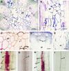
Complementation of the elp1 Mutant Phenotypes by the AtCTL1 Gene. Sections were stained with toluidine blue ([A], [B], [D], and [E]) or phloroglucinol HCl ([C] and [F] to [I]). Lignified cell walls were stained blue by toluidine blue and red by phloroglucinol HCl. (A) Cross-section of an elp1 stem showing aberrant shapes and enlargement of some lignified pith cells. Note that some large pith cells had partial cell walls protruding inward (arrowheads). (B) Longitudinal section of an elp1 stem showing the disorganized arrangement of lignified pith cells. (C) Cross-section of an elp1 stem showing lignin staining in pith cell walls. Note that one large pith cell had partial cell walls protruding inward (arrowheads). (D) and (E) Cross-section (D) and longitudinal section (E) of the stems of elp1 plants transformed with the AtCTL1 gene showing lack of lignin staining in pith cells and the regular shape and arrangement of pith cells. (F) Root of a light-grown elp1 seedling showing lignin staining in endodermal cells. (G) Root of a light-grown elp1 plant transformed with the AtCTL1 gene showing lack of lignin staining in endodermal cells. (H) Hypocotyl of a dark-grown elp1 seedling showing lignin staining in endodermal cells. (I) Hypocotyl of a dark-grown elp1 seedling transformed with the AtCTL1 gene showing lack of lignin staining in endodermal cells. co, cortex; e, epidermis; en, endodermis; pi, pith; x, xylem. Bars in (A) to (E) = 112 μm; bar in (F) = 56 μm for (F) to (I).
Figure 3.
Figure 3.
Structure of the AtCTL1 Gene and Nature of the elp1 Mutation. (A) Exon and intron organization of the AtCTL1 gene. The AtCTL1 gene is 1285 bp long from the start codon (nucleotide 1) to the stop codon (nucleotide 1285). A single nucleotide mutation was found at nucleotide 701 in elp1. Black boxes indicate exons, and lines between exons indicate introns. (B) Effect of the single nucleotide mutation in elp1 on the translation of the predicted protein. Shown are residues of nucleotides and amino acids around the mutation site. The G-to-A transition (asterisk) changes the wild-type codon that encodes a tryptophan residue to a stop codon. (C) Elimination of a ScrF1 site in the mutant elp1 cDNA. The single nucleotide mutation in elp1 happens to occur at a ScrF1 restriction endonuclease cleavage site. This is revealed readily by digesting reverse transcriptase–mediated PCR-amplified cDNA fragments with ScrF1, which shows that the ScrF1 site is missing in the elp1 cDNA compared with the wild-type (WT) AtCTL1 cDNA.
Figure 4.
Figure 4.
Analysis of the Deduced Amino Acid Sequences of AtCTL1. (A) Amino acid sequence alignment of the catalytic regions of AtCTL1 and a few other chitinase-like proteins from Arabidopsis (AtCTL2 and BCHI), potato (PotCHI), tomato (TomCHI), rice (RiCHI), and tobacco (TobCHI). Identical amino acid residues are indicated by colons. Dashed lines are gaps introduced to maximize the identity. The putative secretion signal peptide sequence in AtCTL1 is underlined. Amino acid residues known to be conserved among chitinases are indicated by asterisks. (B) Hydropathy plot of AtCTL1 showing a secretion signal peptide sequence at the N terminus.
Figure 5.
Figure 5.
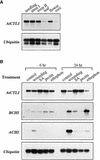
Expression of the AtCTL1 Gene. Total RNA was isolated from different plant organs or stress-treated seedlings and used for reverse transcriptase–mediated PCR analysis. The ubiquitin gene was used as an internal control for PCR. (A) Ubiquitous expression of the AtCTL1 gene in different organs. The seedlings were 3 weeks old. Mature leaves, mature roots, and flowers were from 8-week-old plants. Stem I and stem II were from 4- and 8-week-old plants, respectively. (B) Expression of the AtCTL1, basic chitinase (BCHI), and acidic chitinase (ACHI) genes in response to wounding, salicylic acid (SA), pectin fragments, and ethephon. Three-week-old seedlings were treated with water (control) or other conditions for 6 and 24 hr before being used for expression analysis. The BCHI and ACHI genes were highly induced by various treatments, whereas the gene expression of AtCTL1 was not altered.
Figure 6.
Figure 6.
Morphology of the Wild Type and the elp1 Mutant. (A) and (B) Etiolated seedlings showing decreased length and increased width of hypocotyls of elp1 (B) compared with the wild type (A). (C) and (D) Light-grown seedlings showing a reduction in root length of elp1 (D) compared with the wild type (C). (E) and (F) Visualization of etiolated hypocotyl cells showing a decrease in length of epidermal cells in elp1 (F) compared with the wild type (E). (G) and (H) Close-up of roots of light-grown seedlings showing a dramatic increase in root hair number and length in elp1 (H) compared with the wild type (G). (I) and (J) Visualization of root cells showing a decrease in length of epidermal cells in elp1 (J) compared with the wild type (I). (K) Five-week-old plants showing a reduction in length of inflorescence stems of elp1 (right) compared with the wild type (left). Bars in (A) and (B) = 1.4 mm; bars in (C) and (D) = 1.0 mm; bar in (E) = 80 μm for (E), (F), (I), and (J); bars in (G) and (H) = 0.47 mm.
Figure 7.
Figure 7.
Effects of ACC, Ag+, and AVG on the Morphology of elp1 Seedlings. Four-day-old etiolated seedlings ([A] to [F]) and 5-day-old light-grown seedlings ([G] to [L]) in the absence or presence of ACC (0.1 μM for light-grown seedlings and 5 μM for dark-grown seedlings), Ag+ (1 μM), or AVG (0.5 μM) were used for observation of their morphology. (A) Etiolated hypocotyls of the wild type. (B) Etiolated hypocotyls of the wild type grown in the presence of ACC. (C) Etiolated hypocotyls of elp1 showing increased hook curvature and hypocotyls width compared with the wild type (A). (D) Etiolated hypocotyls of elp1 grown in the presence of ACC showing further increased hook curvature and hypocotyl width. (E) and (F) Etiolated hypocotyls of elp1 grown in the presence of Ag+ (E) and AVG (F) showing decreased hook curvature and hypocotyl width. (G) Roots of light-grown seedlings of the wild type. (H) Roots of the wild-type seedlings grown in the presence of ACC showing increased number of root hairs. (I) Roots of light-grown elp1 seedlings showing increased number of root hairs compared with the wild type (G). (J) Roots of elp1 seedlings grown in the presence of ACC showing increased number of root hairs. (K) and (L) Roots of the elp1 mutant seedlings grown in the presence of Ag+ (K) and AVG (L) showing a dramatic decrease in root hair number and length. Bar in (A) = 0.47 mm for (A) to (L).
Figure 8.
Figure 8.
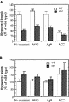
Measurement of the Length and Width of Etiolated Hypocotyls of the Wild Type (WT) and the elp1 Mutant Treated with AVG, Ag+, or ACC. Seed were germinated and grown in the dark for 4 days in the presence of AVG (0.5 μM), Ag+ (1 μM), or ACC (5 μM) before measurement. The length or width of the wild-type hypocotyls without drug treatment were taken as 100, and those of the wild type with drug treatment and all elp1 samples were expressed as percentages of the wild type without drug treatment. Data are means ±se from 20 samples. (A) Hypocotyl length of the wild type and elp1. (B) Hypocotyl width of the wild type and elp1.
Figure 9.
Figure 9.
Measurement of Roots and Root Hairs of Light-Grown Wild-Type (WT) and elp1 Mutant Seedlings Treated with AVG, Ag+, or ACC. Seed were germinated and grown in the light for 5 days in the presence of AVG (0.5 μM), Ag+ (1 μM), or ACC (0.1 μM) before measurement. Parameters of roots or root hairs of the wild type without drug treatment were taken as 100, and those of the wild type with drug treatment and all elp1 samples were expressed as percentages of the wild type without drug treatment. Data are means ±se from 20 samples. (A) and (B) Root length (A) and width (B) of the wild type and elp1. (C) and (D) Root hair number (C) and length (D) of the wild type and elp1.
Figure 10.
Figure 10.
Expression of the Basic Chitinase Gene in the Wild Type (WT) and the elp1 Mutant. Four-day-old etiolated seedlings or 3-week-old light-grown plants were used for total RNA isolation and subsequent reverse transcriptase–mediated PCR analysis of the basic chitinase (BCHI) gene. The ubiquitin gene was used as an internal control for PCR. Although the expression of the BCHI gene was barely detectable in the wild type, it was highly induced in the elp1 mutant.

Similar articles

Cited by

References

    1. Ancillo, G., Witte, B., Schmelzer, E., and Kombrink, E. (1999). A distinct member of the basic (class I) chitinase gene family in potato is specifically expressed in epidermal cells. Plant Mol. Biol. 39, 1137–1151. - PubMed
    1. Bechtold, N., and Bouchez, D. (1994). In planta Agrobacterium-mediated transformation of adult Arabidopsis thaliana plants by vacuum infiltration. In Gene Transfer to Plants, I. Potrykus and G. Spangenberg, eds (Berlin: Springer-Verlag), pp. 19–23.
    1. Benhamou, N., and Asselin, A. (1989). Attempted localization of a substrate for chitinases in plant cells reveals abundant N-acetyl-d-glucosamine residues in secondary walls. Biol. Cell 67, 341–350.
    1. Bent, A.F., Kunkel, B.N., Dahlbeck, D., Brown, K.L., Schmidt, R., Giraudat, J., Leung, J., and Staskawicz, B.J. (1994). RPS2 of Arabidopsis thaliana: A leucine-rich repeat class of plant disease resistance genes. Science 265, 1856–1860. - PubMed
    1. Broglie, K., Chet, I., Hollyday, M., Cressman, R., Biddle, P., Knowlton, S., Mauvais, C.J., and Broglie, R. (1991). Transgenic plants with enhanced resistance to the fungal pathogen Rhizoctonia solani. Science 254, 1194–1197. - PubMed

Publication types

MeSH terms

Associated data