. 2002 Jan;79(1):124-36.
doi: 10.1006/geno.2001.6679.

Sequence analysis of LRPPRC and its SEC1 domain interaction partners suggests roles in cytoskeletal organization, vesicular trafficking, nucleocytosolic shuttling, and chromosome activity

Affiliations

Sequence analysis of LRPPRC and its SEC1 domain interaction partners suggests roles in cytoskeletal organization, vesicular trafficking, nucleocytosolic shuttling, and chromosome activity

Leyuan Liu et al. Genomics. 2002 Jan.

Abstract

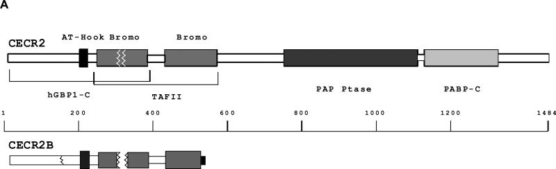
LRPPRC (originally called LRP130) is an intracellular, 130-kD, leucine-rich protein that copurifies with the fibroblast growth factor receptor from liver cell extracts and has been detected in diverse multiprotein complexes from the cell membrane, cytoskeleton, and nucleus. Here we report results of a sequence homology analysis of LRPPRC and its SEC1 domain interactive partners. We found that 23 copies of tandem repeats that are similar to pentatricopeptide, tetratricopeptide, and huntingtin-elongation A subunit-TOR repeats characterize the LRPPRC sequence. The amino terminus exhibits multiple copies of leucine-rich nuclear transport signals followed by ENTH, DUF28, and SEC1 homology domains. We used the SEC1 domain to trap interactive partners expressed from a human liver cDNA library. Interactive C19ORF5 (XP_038600) exhibited a strong homology to microtubule-associated proteins and a potential arginine-rich mRNA binding motif. UXT (XP_033860) exhibited alpha-helical properties homologous to the actin-associated spectrin repeat and L/I heptad repeats in mobile transcription factors. C6ORF34 (XP_004305) was homologous to the non-DNA-binding carboxy terminus of the Escherichia coli Rob transcription factor. CECR2 (AAK15343) exhibited a transcription factor AT-hook motif next to two bromodomains and a homology to guanylatebinding protein-1. Together these features suggest a regulatory role of LRPPRC and its SEC1 domain-interactive partners in integration of cytoskeletal networks with vesicular trafficking, nucleocytosolic shuttling, transcription, chromosome remodeling, and cytokinesis.


Figures

FIG. 1
FIG. 1
Expression of LRPPRC mRNA. Poly(A) mRNA from the indicated tissues was subjected to Northern hybridization analysis with an LRPPRC cDNA spanning the coding region employed in the yeast two-hybrid screen (Trap B, Fig. 2B) and β-actin cDNA as described in Materials and Methods. PBL, peripheral blood leukocytes.
FIG. 2
FIG. 2
Domain structure of LRPPRC predicted from sequence. (A) Homology and secondary structure of the LRP tandem repeats. Repeat sequences and inclusive residues are numbered at right. Residues within an α-helix predicted by PHD are shaded and conserved residues according to type of amino acid are in black. A consensus LRP repeat is indicated showing the most common residue at a conserved site throughout the 23 repeats. Similar, but not identical, residues are indicated in lower case. The consensus, most commonly inter-helical, gly and basic residues (E/D) are underlined. The consensus TPR [6], PPR [6] and HEAT [7] repeat is also indicated. (B) Schematic of the LRP repeat and sequence homology domain structure of LRPPRC.
FIG. 3
FIG. 3
Predicted homologies to ENTH, DUF28 and SEC1 structural domains. Examples and alignments are from the Pfam search. The GenBank entry name of examples is indicated followed by the inclusive range of sequence compared. Residues in black indicate identity and shaded residues indicate amino acids of similar property in respect to hydrophobicity, charge and/or size. (A) ENTH family proteins. (B) DUF28 family proteins. (C) SEC1 family proteins.
FIG. 3
FIG. 3
Predicted homologies to ENTH, DUF28 and SEC1 structural domains. Examples and alignments are from the Pfam search. The GenBank entry name of examples is indicated followed by the inclusive range of sequence compared. Residues in black indicate identity and shaded residues indicate amino acids of similar property in respect to hydrophobicity, charge and/or size. (A) ENTH family proteins. (B) DUF28 family proteins. (C) SEC1 family proteins.
FIG. 4
FIG. 4
Interaction of expression products from cloned human liver cDNAs LRPPRC in the yeast two-hybrid system. (A) Interaction with Trap B containing the SEC1 subdomain of LRPPRC. The five plasmids carrying genes coding for C19ORF5, UXT, C6ORF34B, CECR2B and fibronectin (FN) fused with the activation domain that survived the positive selection series in the yeast two-hybrid complementation system described in the text were co-transformed with the pGBKT7-BD-trap B and full-length LRPPRC fusion construct into yeast AH109 cells. The co-transformants were initially grown on SD/-Leu-Trp plates and re-streaked on QDO plates (Materials and Methods). The β-galactosidase activity in cell lysates was measured spectrophotometrically using the substrate o-nitrophenyl-β-D-galactopyranoside. The data indicated is the mean (± SE) of three independent co-transformations. (B) Interaction of SEC1 interactive substrates with full length LRPPRC. The interaction of the five gene products with the Trap B and entire LRPPRC were compared by ability of co-transformed yeast to grow on QDO plates.
FIG. 5
FIG. 5
Expression of the LRPPRC-interacting partners C19ORF5, UXT, C6ORF34, CECR2 and control β-actin mRNAs in multiple human tissues. Specific probes were described in Materials and Methods. Three CECR2 probes were employed with similar results.
FIG. 6
FIG. 6
Homology of C19ORF5 to microtubule-associated proteins (MAP). (A) Schematic of domain homology to MAP1A (MAPA_RAT). (B) Homology of the N-terminus of C19ORF5. (C) The C-terminus of C19ORF5. The solid bar indicates a potential RNA binding motif. The indicated alignment was based on the MAXHOM multiple sequence alignment program. The GenBank entry name of examples is indicated followed by the inclusive range of sequence compared. Residues in black indicate identity and shaded residues indicate amino acids of similar property in respect to hydrophobicity, charge and/or size. Identical and similar residues are bolded or shaded, respectively. Two lowercase residues indicate an omission of a stretch of sequence with insignificant homology between the residues.
FIG. 7
FIG. 7
Examples of UXT homology to spectrin repeat-containing proteins and two membrane-bound transcription factors. (A) Spectrin repeat proteins. Alignment to 954-residue human dystrophin-related protein 2 (DRP2) was based on the MAXHOM multiple sequence alignment program, while that to 288-residue syntaxin 1a and 444-residue cortexillin I was predictions of structural homology based on sequence using the 3D-PSSM program. (B, C) Mobile transcription factors SREB1 and STAT3b. Full-length SREB1 and STAT3b consist of 1147 and 770 residues, respectively. Alignment was also based on the 3D-PSSM program. The signature tyr-335 in the basic region of SREB1 is noted by triangle and the L/I residues of the four heptad repeats in UXT are noted by solid circle. Residue identity and similarities are indicated in black and gray, respectively. The secondary structure (C, loop; E, β-strand; and H, α-helix) to which each residue is predicted to contribute is indicated above each residue. The actual secondary structure from x-ray or NMR for each homologue is indicated below each residue
FIG. 8
FIG. 8
Homology of C6ORF34 to the regulatory domain of the E. coli transcriptional factor Rob. The alignment is based on the 3D-PSSM analysis with the predicted and actual structures indicated as described in Fig. 7.
FIG. 9
FIG. 9
Homology domains of the CECR2 protein. (A) Schematic of the homology domain structure. The indicated homology domains are shown in proportion to the full-length 1484-residue CECR2 protein at top. The deletion in the bromodomain I of CECR2 due to exon skipping is indicated (white wavy lines). The black wavy line at left in CECR2B indicates the point of N-terminal fusion with the library expression vector. (B) GBP1 and the AT-Hook Homology. Alignment to the C-terminus of 592-residue human GBP1 is based on the 3D-PSSM analysis with the predicted and actual structures indicated as described in Fig. 7. The AT-Hook sequence is overlined, its highly conserved signature GRP residues are underlined and bromodomain I is indicated. The deletion which results from exon skipping is between the solid triangles. (C) Example of homology to a two-bromodomain transcription factor. Both Pfam and 3D-PSSM searches detected the TFIID homologue because of its tandem bromodomains (overlined with bar). Alignment was performed with 3D-PSSM analysis. The point at which the alternate exon in CECR2 begins is indicated by the open triangle. and the C-terminus with a star. (D) Structural domain homology with PABP-C. Alignment to the C-terminus of 636-residue human poly A binding protein 1 was also based on the 3D-PSSM search. Residue identity and similarities predicted and actual secondary structure are indicated as described in Fig. 7.
FIG. 9
FIG. 9
Homology domains of the CECR2 protein. (A) Schematic of the homology domain structure. The indicated homology domains are shown in proportion to the full-length 1484-residue CECR2 protein at top. The deletion in the bromodomain I of CECR2 due to exon skipping is indicated (white wavy lines). The black wavy line at left in CECR2B indicates the point of N-terminal fusion with the library expression vector. (B) GBP1 and the AT-Hook Homology. Alignment to the C-terminus of 592-residue human GBP1 is based on the 3D-PSSM analysis with the predicted and actual structures indicated as described in Fig. 7. The AT-Hook sequence is overlined, its highly conserved signature GRP residues are underlined and bromodomain I is indicated. The deletion which results from exon skipping is between the solid triangles. (C) Example of homology to a two-bromodomain transcription factor. Both Pfam and 3D-PSSM searches detected the TFIID homologue because of its tandem bromodomains (overlined with bar). Alignment was performed with 3D-PSSM analysis. The point at which the alternate exon in CECR2 begins is indicated by the open triangle. and the C-terminus with a star. (D) Structural domain homology with PABP-C. Alignment to the C-terminus of 636-residue human poly A binding protein 1 was also based on the 3D-PSSM search. Residue identity and similarities predicted and actual secondary structure are indicated as described in Fig. 7.

References

    1. DiSorbo D, Shi EG, McKeehan WL. Purification form human hepatoma cells of a 130-kDa membrane glycoprotein with properties of the heparin-binding growth factor receptor. Biochem. Biophys. Res. Commun. 1988;157:1007–1014. - PubMed
    1. Hou J, Wang F, McKeehan WL. Molecular cloning and expression of the gene for a major leucine-rich protein from human hepatoblastoma cells (HepG2). In Vitro Cell. Dev. Biol. Anim. 1994;30A:111–114. - PubMed
    1. Ghiso NS, Lennon GG. LRP130 gene assigned to chromosome 2. In Vitro Cell. Dev. Biol. Anim. 1994;30A:744. - PubMed
    1. Suzuki Y, Wanaka A, Tohyama M, Takagi T. Identification of differentially expressed mRNAs during neuronal differentiation of P19 embryonal carcinoma cells. Neurosci. Res. 1995;23:65–71. - PubMed
    1. Kan M, et al. High and low affinity binding of heparin-binding growth factor to a 130-kDa receptor correlates with stimulation and inhibition of growth of a differentiated human hepatoma cell. J. Biol. Chem. 1988;263:11306–11313. - PubMed

Associated data