

MR analysis of the substantia innominata in normal aging, Alzheimer disease, and other types of dementia
- PMID: 11827872
- PMCID: PMC7975505
MR analysis of the substantia innominata in normal aging, Alzheimer disease, and other types of dementia
Abstract
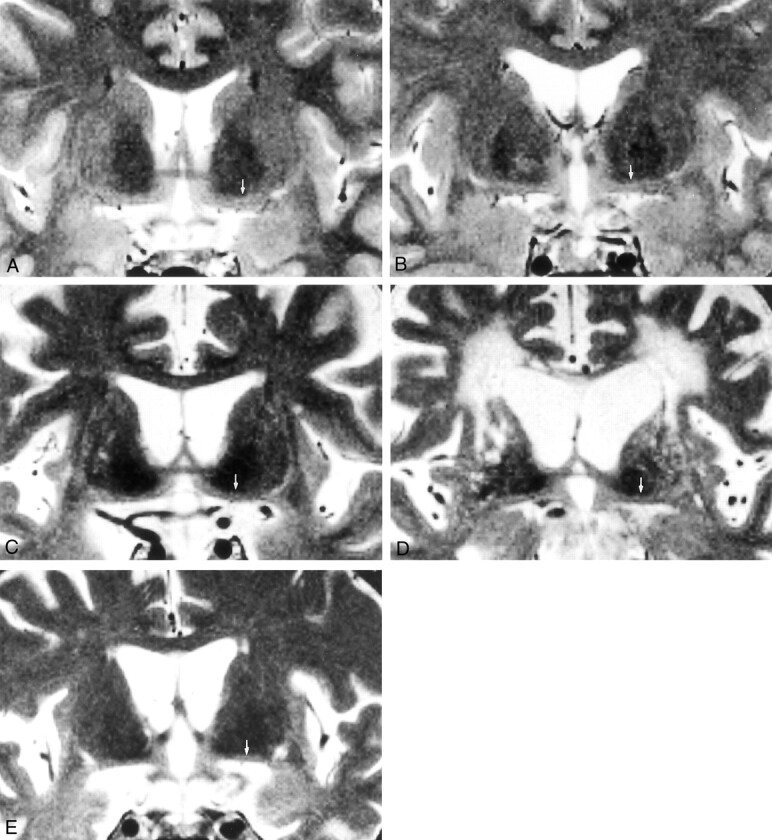
Background and purpose: The substantia innominata can be visualized on coronal thin-section T2-weighted MR images. The purpose of this study was to investigate the morphologic changes of the substantia innominata in normal aging by using MR imaging and to determine whether the changes in this structure on MR images were specific to Alzheimer disease (AD).
Methods: The thickness of the substantia innominata was measured on the coronal T2-weighted image obtained through the anterior commissure in 39 healthy control subjects (age range, 25-86 y; mean age, 62 y); 39 patients with AD; and 36 patients with non-AD dementia, including vascular dementia, frontotemporal dementia, and Parkinson disease with dementia.
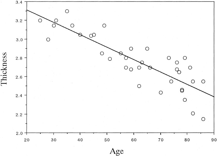
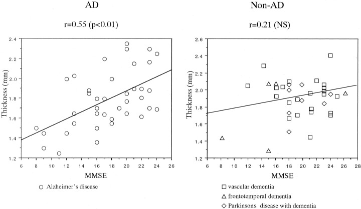
Results: In the control subjects, the thickness of the substantia innominata significantly decreased with age. Compared with age-matched control subjects, both patients with AD and patients with non-AD dementia had significant atrophy of the substantia innominata. The thickness of the substantia innominata significantly correlated with scores from the Mini-Mental State Examination in patients with AD but not in patients with non-AD dementia.
Conclusion: MR analysis reveals age-related shrinkage of the substantia innominata. Atrophy of the substantia innominata, which reflects degeneration in the nucleus basalis of Meynert, is pronounced both in patients with AD and in those with non-AD dementia. MR imaging features in this structure may not be specific to AD.
Figures
Comment in
-
Past glory and future promise: maximizing and improving understanding of atrophy patterns in the diagnosis of degenerative dementias.AJNR Am J Neuroradiol. 2002 Jan;23(1):33-4. AJNR Am J Neuroradiol. 2002. PMID: 11827873 Free PMC article. No abstract available.
References
-
- Whitehouse PJ, Price DL, Clark AW, Coyle JT, DeLong MR. Alzheimer disease: evidence for selective loss of cholinergic neurons in the nucleus basalis. Ann Neurol 1981;10:122–126 - PubMed
-
- Whitehouse PJ, Price DL, Struble RG, Clark AW, Coyle JT, DeLong MR. Alzheimer’s disease and senile dementia: loss of neurons in the basal forebrain. Science 1982;215:1237–1239 - PubMed
-
- McKhann G, Drachman D, Folstein M, Katzman R, Price D, Stadlan EM. Clinical diagnosis of Alzheimer’s disease: report of the NINCDS-ADRDA Work Group under the auspices of Department of Health and Human Services Task Force on Alzheimer’s disease. Neurology 1984;34:939–944 - PubMed
-
- Hachinski VC, Iliff LD, Zilhka E, et al Cerebral blood flow in dementia. Arch Neurol 1975;32:632–637 - PubMed
MeSH terms
LinkOut - more resources
Full Text Sources
Medical