. 2002 Jan;23(1):67-75.

High-resolution line scan diffusion tensor MR imaging of white matter fiber tract anatomy

Affiliations

High-resolution line scan diffusion tensor MR imaging of white matter fiber tract anatomy

Hatsuho Mamata et al. AJNR Am J Neuroradiol. 2002 Jan.

Abstract

Background and purpose: MR diffusion tensor imaging permits detailed visualization of white matter fiber tracts. This technique, unlike T2-weighted imaging, also provides information about fiber direction. We present findings of normal white matter fiber tract anatomy at high resolution obtained by using line scan diffusion tensor imaging.

Methods: Diffusion tensor images in axial, coronal, and sagittal sections covering the entire brain volume were obtained with line scan diffusion imaging in six healthy volunteers. Images were acquired for b factors 5 and 1000 s/mm(2) at an imaging resolution of 1.7 x 1.7 x 4 mm. For selected regions, images were obtained at a reduced field of view with a spatial resolution of 0.9 x 0.9 x 3 mm. For each pixel, the direction of maximum diffusivity was computed and used to display the course of white matter fibers.

Results: Fiber directions derived from diffusion tensor imaging were consistent with known white matter fiber anatomy. The principal fiber tracts were well observed in all cases. The tracts that were visualized included the following: the arcuate fasciculus; superior and inferior longitudinal fasciculus; uncinate fasciculus; cingulum; external and extreme capsule; internal capsule; corona radiata; auditory and optic radiation; anterior commissure; corpus callosum; pyramidal tract; gracile and cuneatus fasciculus; medial longitudinal fasciculus; rubrospinal, tectospinal, central tegmental, and dorsal trigeminothalamic tract; superior, inferior, and middle cerebellar peduncle; pallidonigral and strionigral fibers; and root fibers of the oculomotor and trigeminal nerve.

Conclusion: We obtained a complete set of detailed white matter fiber anatomy maps of the normal brain by means of line scan diffusion tensor imaging at high resolution. Near large bone structures, line scan produces images with minimal susceptibility artifacts.


Figures
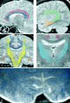
F<sc>ig</sc> 1.
Fig 1.
When viewed in multiple sections, the appearance of the association fibers on the principal eigenvector maps is consistent with known anatomy. A, Sagittal 110 × 110-mm subimage close to the midline, passing through the cingulum (pink). Note that the fibers of the corpus callosum run at a slight angle through the plane (blue dots with short lines). B, More lateral sagittal section obtained through the arcuate fasciculus (green), inferior longitudinal fasciculus (blue), and uncinate fasciculus (orange). C, Coronal section obtained at the level of the posterior limb of the internal capsule. Fibers of the cingulum (pink), arcuate fasciculus (green), and uncinate fasciculus (orange) pass through the section (dots). Fibers of the posterior limb of the internal capsule (yellow lines) are running in-plane. D, High-resolution 55 × 55-mm coronal subimage of the thalamic region. The strionigral and pallidonigral fibers are shown in light blue. E, High-resolution axial surface coil image of the calcarine area at a 120 × 60-mm field of view. U-fibers connecting visual cortices are well seen.
F<sc>ig</sc> 2.
Fig 2.
Subimages (110 × 110-mm field of view) of the projection fibers. A, Lateral sagittal section passing through the corona radiata (yellow lines). B, Axial section obtained through the genu and splenium of the corpus callosum. Corona radiata fibers within the posterior limb (yellow) of the internal capsule can be distinguished by their strong through-plane component (dots), whereas fibers within the anterior limb (orange) of the corpus callosum run in-plane (lines). C, Axial section obtained at the level of the basal ganglia. Fibers in the anterior limb of the internal capsule (orange) are running in-plane (lines), whereas in the posterior limb of the internal capsule (yellow), the fibers run through-plane (dots). Parts of the optic (pink) and auditory (green) radiation can also be seen. D, Coronal section obtained through the trigone of the lateral ventricles. Pink dots indicate through-plane fibers that are part of the optic radiation.
F<sc>ig</sc> 3.
Fig 3.
Commissural fibers of the corpus callosum are observed in sections obtained near the lateral ventricle. A, Coronal section obtained through the insula (110 × 110-mm subimage). In this section, the cingulum (pink) and fibers (blue) interconnecting the frontoparietal cortices of each hemisphere with the corpus callosum can be seen. The fibers of the anterior commissure (green) radiate into the temporal lobes. B, High resolution coronal section (55 × 55-mm subimage) obtained at the level of the anterior commissure (green).
F<sc>ig</sc> 4.
Fig 4.
Subimages (55 × 55-mm field of view) of the brain stem. A, Axial section obtained at the level of the upper midbrain. Very fine fibers that originate from the oculomotor nucleus run through and around the red nucleus and converge to the roots of the oculomotor nerves (green). Note that fibers of the cerebral peduncles are running at a slight angle through the plane (yellow dots with lines). B, Axial section obtained at the level of the lower midbrain. The arrow indicates the decussation of the superior cerebellar peduncle. C, Axial section (110 × 110-mm field of view) obtained at the level of the midbrain-cerebrum junction. The fibers of the optic tract (arrows) connect to the lateral geniculate bodies. Fibers of the optic radiation (pink) pass lateral to the optic tract. D, Axial section obtained at the level of the pons, through the roots of the trigeminal nerve (green). Fibers of the craniocaudal tracts (superior cerebellar peduncle, medial longitudinal fasciculus, tectospinal tract, dorsal and ventral trigeminothalamic tracts, central tegmental tract, spinothalamic tract, and rubrospinal tract) can be seen in the posterior part of the pons (pink). The yellow dots indicate fibers of the pyramidal and the frontopontine tracts.
F<sc>ig</sc> 5.
Fig 5.
Subimages of the brain stem. A, Coronal section (110 × 55-mm subimage) of the brain stem obtained at the level of the motor decussation. Fibers of the motor tract cross to the contralateral side at the level of the lower medulla. B, Axial subimage (27.5 × 27.5-mm field of view) of the medulla obtained at the level of the motor decussation. Fibers of the motor tract cross to the contralateral side. Short lines with blue dots indicate low anisotropy. C, Midsagittal subimage (55 × 55-mm field of view) of the brain stem. The pyramidal tract is shown in yellow and the medial longitudinal fasciculus in pink. D, Sagittal section (55 × 55-mm field of view subimage) of the brain stem at a lateral position. Fibers of the pyramidal tract (yellow) enter the pons. The arrowheads indicate fine fibers of the sensory decussation (internal arcuate fibers), running in an anteroposterior direction.
F<sc>ig</sc> 6.
Fig 6.
Subimages (55 × 55-mm field of view) of the cerebellum. A, Coronal section obtained at the level of the fourth ventricle floor. Fibers of the superior (green) and inferior (pink) cerebellar peduncles pass in a craniocaudal direction toward the cerebellar hemisphere, and fibers of the middle cerebellar peduncle (yellow) pass at an oblique angle through the section (dots with lines). B, More posterior coronal section of the cerebellum. Fibers from the cerebellar peduncles branch out toward the cerebellar cortices.

Comment in

References

    1. Ting YL, Bendel P. Thin-section MR imaging of rat brain at 4.7T. J Magn Reson Imaging 1992;2:393–399 - PubMed
    1. Ghosh P, O’Dell M, Narasimhan PT, Fraser SE, Jacobs RE. Mouse lemur microscopic MRI brain atlas. Neuroimage 1994;1:345–349 - PubMed
    1. De Coene B, Hajnal JV, Pennock JM, Bydder GM. MRI of the brain stem using fluid attenuated inversion recovery pulse sequences. Neuroradiology 1993;35:327–331 - PubMed
    1. Wolff SD, Balaban RS. Magnetization transfer contrast (MTC) and tissue water proton relaxation in vivo. Magn Reson Med 1993;29:77–83 - PubMed
    1. Balaban RS, Ceckler TL. Magnetization transfer contrast in magnetic resonance imaging. Magn Reson Q 1992;8:116–137 - PubMed

Publication types