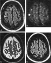
Diffusion-weighted MR imaging in the acute phase of transient ischemic attacks
- PMID: 11827878
- PMCID: PMC7975510
Diffusion-weighted MR imaging in the acute phase of transient ischemic attacks
Abstract
Background and purpose: Radiologic assessment of acute transient ischemic attacks (TIAs) has been handicapped by the low sensitivity of CT and conventional MR imaging for acute small-vessel infarction and the difficulty in differentiating between acute and chronic lesions by use of these methods. Our purpose was to evaluate the incidence of TIA-related infarction by using diffusion-weighted MR imaging to determine whether the presence of a diffusion imaging abnormality correlates with the duration of symptoms or cause of TIA.
Methods: We prospectively studied 58 consecutive patients with acute TIA by use of diffusion-weighted imaging. All MR imaging was performed with a 1.5-T whole-body system with 24-mT/m gradient strength and an echo-planar-capable receiver. All patients were imaged within 10 days of stroke onset.
Results: Thirty-nine patients (67%) manifested a diffusion imaging abnormality consistent with acute ischemia. Cortical lesions were identified in 54% of these patients; most of them associated with other acute ischemic lesions. Subcortical lesions were identified in 46%; most of them were isolated from other lesions. The mean duration of symptoms in patients with no TIA-related diffusion imaging abnormalities was 0.96 hours (median, 0.33 hours) compared with a mean of 6.85 hours (median, 1.53 hours) in patients with diffusion imaging abnormalities (P =.025, Mann-Whitney U test). This significant correlation between the duration of TIA symptoms and the presence of TIA-related abnormalities was lost when we excluded from the analysis patients whose symptoms lasted longer than 6 hours (P =.513, Mann-Whitney U test). No significant correlation was observed between the size of TIA-related lesions and the duration of symptoms or cause of TIA.
Conclusion: Two thirds of our TIA patients showed focal abnormalities indicative of acute ischemic lesions on diffusion-weighted images. This incidence is higher than that previously reported in the literature. The presence of such abnormalities increased with increasing total symptom duration, but this relation was not observed when only patients whose symptoms lasted less than 6 hours were considered. No significant correlation was observed between the cause and presence of TIA-related lesions on diffusion-weighted MR images. These TIA-related lesions are probably irreversible and may lead to subsequent infarct.
Figures


Comment in
-
Transient ischemic attacks: added specificity from modern MR imaging.AJNR Am J Neuroradiol. 2002 Jan;23(1):4-5. AJNR Am J Neuroradiol. 2002. PMID: 11827867 Free PMC article. No abstract available.
References
-
- The Ad Hoc Committee on the Classification and Outline of Cerebrovascular Disease, II. Stroke 1975;6:566–616 - PubMed
-
- Dyken MI, Conneally M, Haerer AF, et al. Cooperative study of hospital frequency and character of transient ischemic attacks, I: background, organization and clinical surgery. JAMA 1977;237:882–886 - PubMed
-
- Dennis M, Bamford J, Sandercock P, Warlow C. Prognosis of transient ischemic attacks in the Oxfordshire Community Stroke Project. Stroke 1990;21:848–853 - PubMed
-
- Streiffler JY, Benavente OR, Harbison JW, Eliasziw M, Hachinski VC, Barnett HJM. Prognostic implications of retinal versus hemispheric TIA in patients with high grade carotid stenosis: observations from NASCET (abstract). Stroke 1992;23:159
-
- Toole JF. The Willis lecture: transient ischemic attacks, scientific method, and new realities. Stroke 1983;14:433–437 - PubMed
MeSH terms
LinkOut - more resources
Full Text Sources
Medical
