. 2002 Jan;23(1):143-50.

Analysis of the cerebral cortex in holoprosencephaly with attention to the sylvian fissures

Affiliations

Analysis of the cerebral cortex in holoprosencephaly with attention to the sylvian fissures

A James Barkovich et al. AJNR Am J Neuroradiol. 2002 Jan.

Abstract

Background and purpose: Analysis of specific features in the brain of patients with holoprosencephaly (HPE) may clarify normal and abnormal brain development and help predict outcomes for specific children. We assessed sulcal and gyral patterns of cerebral cortex in patients with HPE and developed a method of grading brain development.

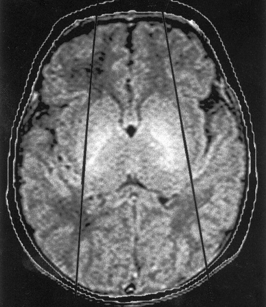
Methods: Neuroimaging studies (75 MR imaging, 21 CT) of 96 patients with HPE were retrospectively reviewed, with specific attention paid to the cerebral cortex. Thickness of cortex, width of gyri, and depth of sulci were assessed subjectively and by measurement. The angle between lines drawn tangential to the sylvian fissures ("sylvian angle") was measured in each patient with HPE and in 20 control patients.

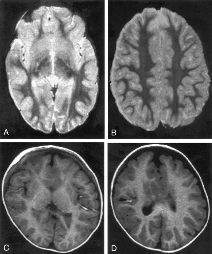
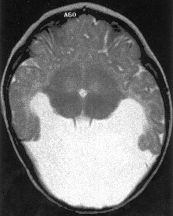
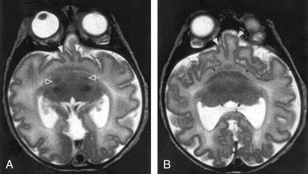
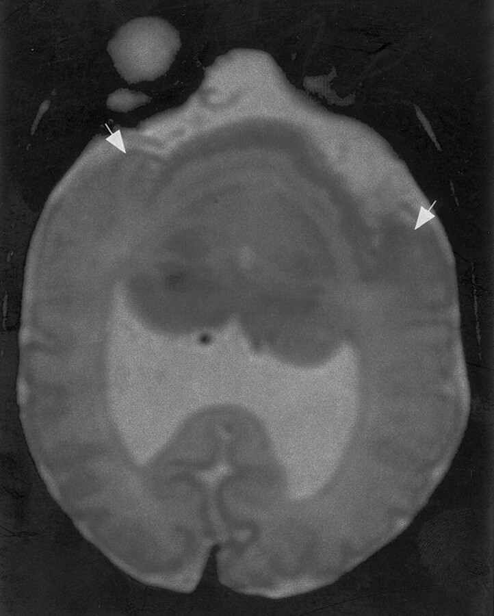
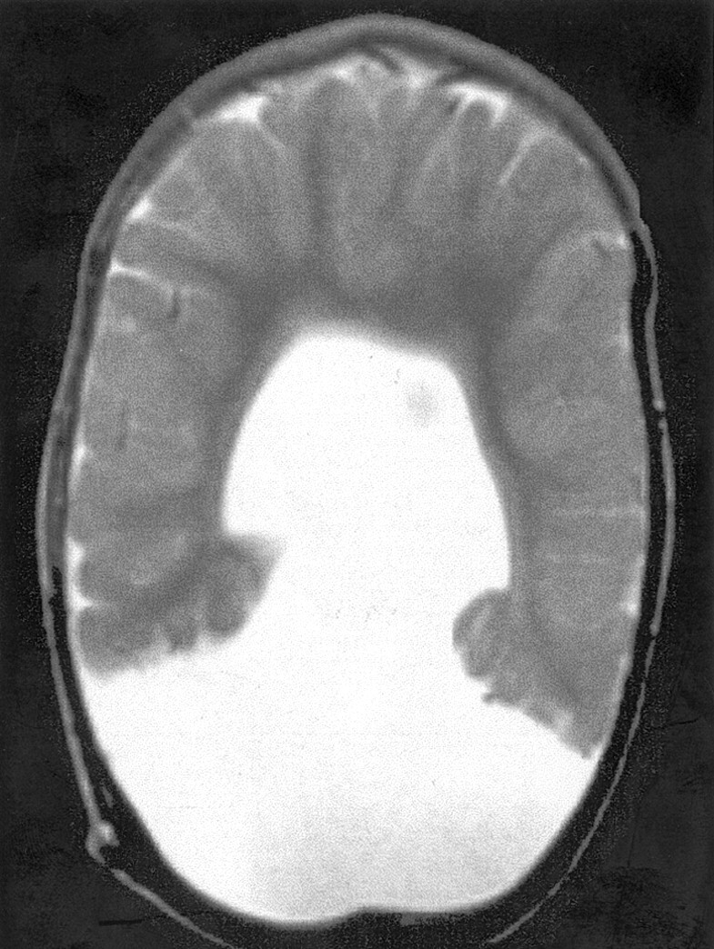
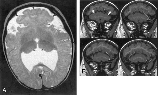
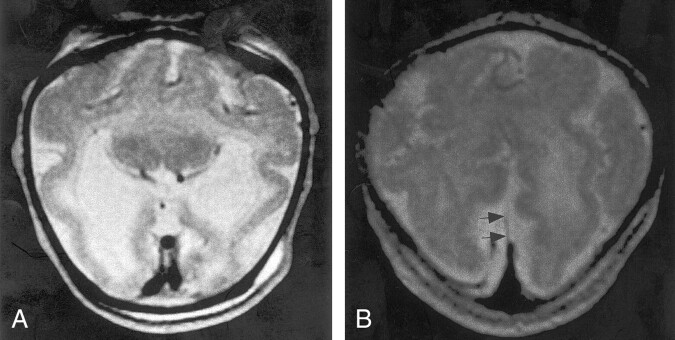
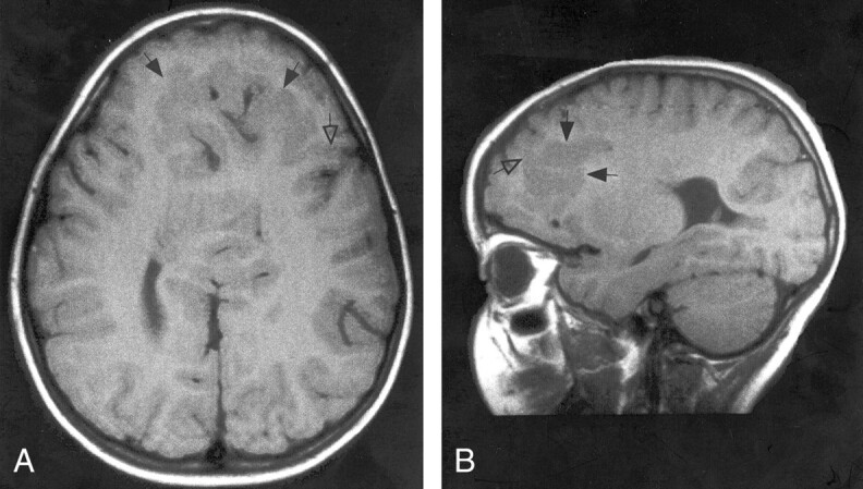
Results: Thickness of cortex was normal in all 96 patients. Gyral shape and width and sulcal depth were normal in 80 patients. Twelve patients, all with very severe HPE and microcephaly, had reduced sulcal depth, diffusely in eight and limited to the anteromedial cortex in four with lobar HPE. Four patients had subcortical heterotopia, located anterior to the interhemispheric fissure, associated with shallow sulci in the overlying cortex. Sylvian fissures were displaced further anteriorly and medially as HPE became more severe, until, in the most severe cases, no sylvian fissures could be identified. Sylvian angle measurements corresponded closely with severity of HPE, being largest in the most severe and smallest in the least severe cases. All patients with HPE had sylvian angles significantly larger than the mean of 15 degrees measured in the control patients.

Conclusion: The only true malformations of cortical development were subcortical heterotopia. However, diffuse and focal abnormal sulci were observed. We propose our sylvian angle measurement of extent of frontal lobe development as an objective means of quantifying the severity of HPE.


Figures

F<sc>ig</sc> 1.
Fig 1.
Axial spin-echo (3000/120 [TR/TE]) MR image shows the sylvian angle in a healthy neonate. Black lines are drawn tangential to the sylvian fissures at the level of the basal ganglia. The anterior angle is the sylvian angle.
F<sc>ig</sc> 2.
Fig 2.
MR images show two cases of mild HPE. Both cases were classified as lobar HPE and have sylvian angles of 25°. Both have small, dysplastic frontal horns and both have a large amount of brain tissue anterior to the sylvian fissures. A and B, Spin-echo (2500/120) MR images show normal-appearing sulci and gyri in the posterior two-thirds. C and D, Inversion-recovery (2000/800/inversion time, 12 ms) MR images show normal-appearing sulci and gyri in the posterior half. The medial frontal sulci appear dysplastic.
F<sc>ig</sc> 3.
Fig 3.
Spin-echo (3000/120) MR image shows HPE of moderate severity, classified as less dysplastic semilobar HPE. The sylvian angle is 95°, and little brain tissue lies anterior to the sylvian fissures.
F<sc>ig</sc> 4.
Fig 4.
A and B, Spin-echo (3000/120) MR images show HPE of moderate severity, classified as less dysplastic semilobar HPE. The sylvian angle is 110°. Only two gyri (solid white arrows) are positioned between the sylvian fissures anteriorly. The basal ganglia are less well developed than those shown in Figure 3, being only a crescent of gray matter anterior to the thalami in B. A thin curvilinear area of gray matter hypointensity (small black arrows, B) is seen between the basal ganglia and presumed insula; this is thought to represent claustrum. Two foci of hypointensity (open white arrows, A) are seen posterior to the basal ganglia crescent but anterior to the thalami in A. These are thought to represent myelinated white matter in the internal capsules.
F<sc>ig</sc> 5.
Fig 5.
Fast spin-echo (3600/95) MR image shows HPE of moderate severity, classified as more dysplastic semilobar HPE. The anteriormost portion of the cerebrum appears to be a single continuous sylvian fissure. This fissure appears to be composed of the posterior halves of two sylvian fissures, with bilateral posterior opercula (arrows) forming the lateral borders, as if the anterior halves of the fissures had never formed and the posterior halves merged together.
F<sc>ig</sc> 6.
Fig 6.
Spin-echo (2500/70) MR image shows severe HPE. No sylvian fissures are identified and, thus, no sylvian angle measured. Gyri have normal, uniform width, and sulci have normal, uniform depth. No specific gyri or sulci are identified.
F<sc>ig</sc> 7.
Fig 7.
MR images show HPE of moderate severity, classified as less dysplastic semilobar. The sylvian angle is 55°. A, Axial fast spin-echo (4000/84) MR image shows that a mushroom-shaped region of brain (arrowheads) grows anteriorly from the region between the anterior aspects of the sylvian fissures. B, Coronal spoiled gradient-recalled acquisition in a steady state (36/13) MR images show the large mushroom-shaped region (arrows, B) lying below the remainder of the cerebrum in the most anterior aspect of the calvaria.
F<sc>ig</sc> 8.
Fig 8.
MR images show severe HPE, classified as alobar, with sylvian angle of 135°. First impression of the cerebral cortex is one of pachygyria, but the cortical thickness measures only 3 mm. The cortex appears thick because of the profound microcephaly. This case illustrates some of the difficulties of the DeMyer classification, as the interhemispheric fissure and ventricular systems are much better developed than the basal ganglia and white matter. The basal ganglia, might be considered alobar HPE, but the interhemispheric development suggests mild semilobar HPE. A, Axial fast spin-echo (4000/90) MR image shows apparently thickened cortex with few gyri and shallow sulci. Thalami are incompletely separated, and no basal ganglia are seen, nor were they seen at other levels. B, Axial fast spin-echo (4000/90) MR image obtained at a higher level shows an interhemispheric fissure spanning the posterior two-thirds of the cerebrum; the falx cerebri (arrows) is present posteriorly.
F<sc>ig</sc> 9.
Fig 9.
A and B, Spin-echo (700/16) MR images show mild HPE, classified as lobar, with a sylvian angle of 25°. Axial (A) and sagittal (B) views show curvilinear subcortical heterotopia (solid arrows) in the anterior cerebrum. The heterotopia connect with the overlying cortex in several places (open arrow). The overlying cortex is thin with shallow sulci.

References

    1. DeMyer W. Holoprosencephaly (cyclopia-arhinencephaly). In: Myrianthopoulos N, ed. Malformations. New York: Elsevier;1987. :225–244
    1. DeMyer W, Zeman W. Alobar holoprosencephaly (arhinencephaly) with median cleft lip and palate: clinical, nosologic, and electroencephalographic considerations. Confin Neurol 1963;23:1–36 - PubMed
    1. DeMyer W, Zeman W, Palmer CG. The face predicts the brain: diagnostic significance of median facial anomalies for holoprosencephaly (arhinencephaly). Pediatrics 1964;34:256–263 - PubMed
    1. Probst FP. The Prosencephalies: Morphology, Neuroradiological Appearances and Differential Diagnosis. Berlin: Springer-Verlag; 1979:46
    1. Golden JA. Towards a greater understanding of the pathogenesis of holoprosencephaly. Brain Dev 1999;21:513–521 - PubMed

Publication types