. 2002 Feb 4;156(3):453-65.
doi: 10.1083/jcb.200111025. Epub 2002 Feb 4.

The yeast protein kinase Mps1p is required for assembly of the integral spindle pole body component Spc42p

Affiliations

The yeast protein kinase Mps1p is required for assembly of the integral spindle pole body component Spc42p

Andrea R Castillo et al. J Cell Biol. .

Abstract

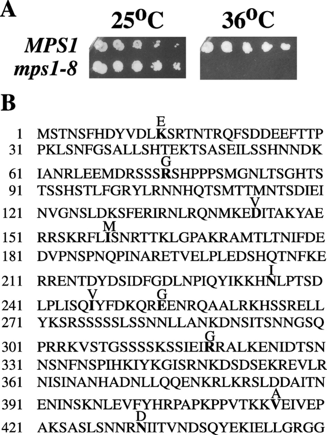
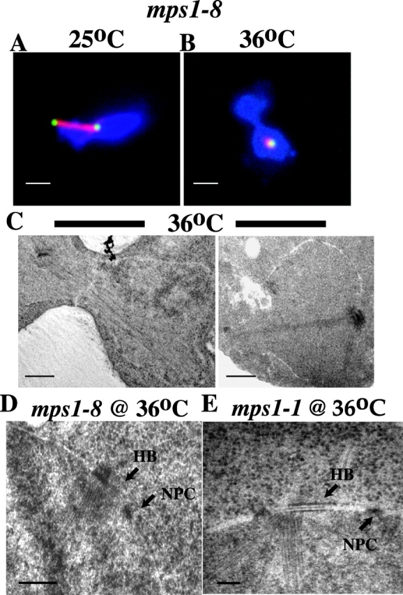
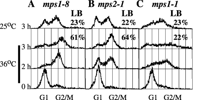
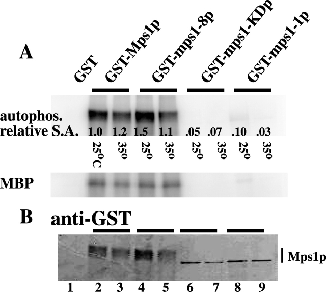
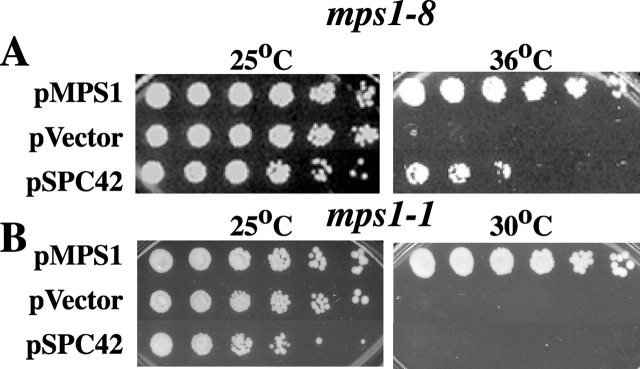
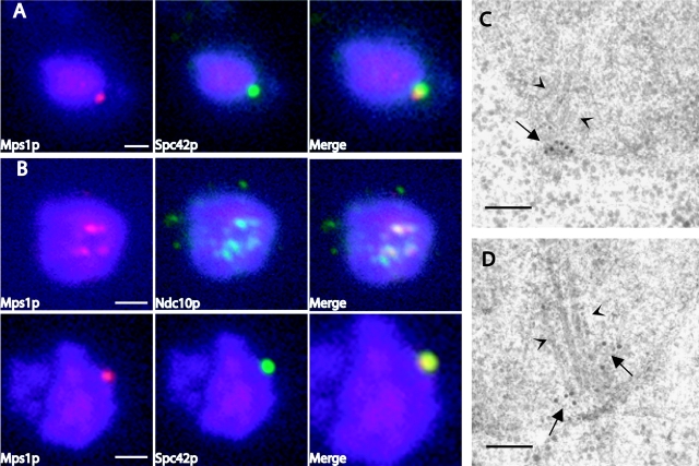
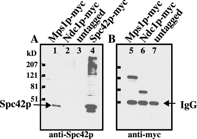
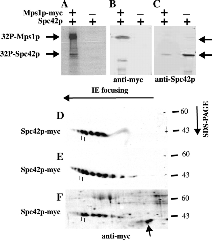
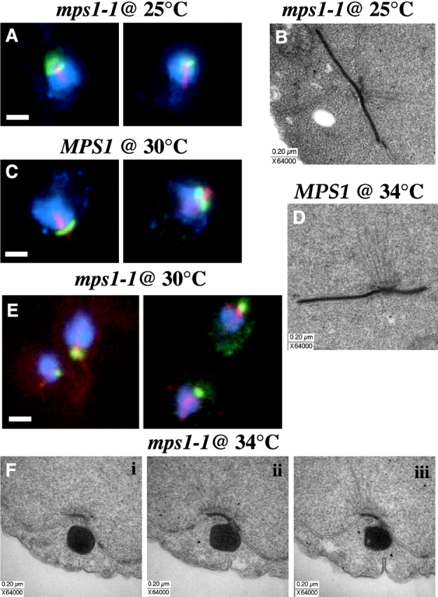
Saccharomyces cerevisiae MPS1 encodes an essential protein kinase that has roles in spindle pole body (SPB) duplication and the spindle checkpoint. Previously characterized MPS1 mutants fail in both functions, leading to aberrant DNA segregation with lethal consequences. Here, we report the identification of a unique conditional allele, mps1-8, that is defective in SPB duplication but not the spindle checkpoint. The mutations in mps1-8 are in the noncatalytic region of MPS1, and analysis of the mutant protein indicates that Mps1-8p has wild-type kinase activity in vitro. A screen for dosage suppressors of the mps1-8 conditional growth phenotype identified the gene encoding the integral SPB component SPC42. Additional analysis revealed that mps1-8 exhibits synthetic growth defects when combined with certain mutant alleles of SPC42. An epitope-tagged version of Mps1p (Mps1p-myc) localizes to SPBs and kinetochores by immunofluorescence microscopy and immuno-EM analysis. This is consistent with the physical interaction we detect between Mps1p and Spc42p by coimmunoprecipitation. Spc42p is a substrate for Mps1p phosphorylation in vitro, and Spc42p phosphorylation is dependent on Mps1p in vivo. Finally, Spc42p assembly is abnormal in a mps1-1 mutant strain. We conclude that Mps1p regulates assembly of the integral SPB component Spc42p during SPB duplication.


Figures

Figure 1.
Figure 1.
A new MPS1 allele, mps1-8 , contains mutations in the noncatalytic region that result in conditional growth at 36°C. (A) A wild-type (WX257-14c) and mps1-8 (ACY54-9b) strain was grown to saturation at 25°C and plated in fivefold serial dilution on rich media. These plates were incubated at 25 or 36°C for 4 d. The mps1-8 strains fail to grow at the restrictive temperature of 36°C. (B) Sequencing of the noncatalytic region (amino acids 1-450) of mps1-8 revealed 10 point mutations that resulted in amino acid changes (K13E, R74G, D143V, I158M, N235I, I244V, E254G, R319G, V415A, N429D).
Figure 2.
Figure 2.
mps1-8 cells fail in SPB duplication when grown at their restrictive temperature (36°C). (A and B) Immunofluorescence images of a mps1-8 (ACY71-14b) strain grown at the permissive (A, 25°C) or restrictive (B, 36°C) temperature for 3 h after release from an α-factor–induced G1 arrest. These cells were fixed and stained with DAPI to visualize DNA (blue), an antibody against α-tubulin to visualize microtubules (red), and Spc42-GFP in this strain (ACY71-14b) identifies the SPBs (green). (A) In mps1-8 cells grown at the permissive temperature (25°C), two foci of Spc42-GFP in the large budded cell indicates that the SPB has duplicated. In this image, a short spindle can be seen between the two SPBs. (B) At the restrictive temperature (36°C), only a single foci of GFP signal is observed in large budded cells (92%, n = 42), indicating SPB duplication has failed to occur. (C–E) Electron micrographs of asynchronously growing mps1-8 (ACY66-4) and mps1-1 (WX241-3b) diploids shifted to the restrictive temperature for 5 h. An unduplicated SPB was detected in serial sections of large budded mps1-8 and mps1-1 cells. Only one section is shown here. The unduplicated SPB of mps1-8 cells (D; n = 17) is associated with half-bridge (HB, arrow) material but lacks the extended half-bridge typical of mps1-1 mutants (E, HB, arrow). NPC, nuclear pore complex. Bars: (A and B) 2.0 μm; (C) 0.4 μm; (D) 0.2 μm; (E) 0.1 μm.
Figure 3.
Figure 3.
mps1-8 cells arrest in mitosis through activation of the spindle checkpoint. Asynchronously growing mps1-8 (ACY54-9b), mps2-1 (SMY-1b), and mps1-1 (WX241-10c) strains were arrested in G1 using α-factor and then released at both permissive (25°C) and restrictive (36°C) temperatures. Samples for flow cytometry were collected at T = 0 (G1 arrest), 2, and 3 h after release from the G1 arrest (only the 3-h time point shown for cells at 25°C). (A) The mitotic arrest observed in mps1-8 cells at the restrictive temperature is seen as the accumulation of cells with a G2 DNA content and large budded (LB) cell morphology (61%). At the permissive temperature, mps1-8 cells return to cycling asynchronously. (B) For comparison, mps2-1 cells also arrest in mitosis after they fail in SPB duplication at the restrictive temperature. (C) A mps1-1 strain serves as a negative control for a mutant that fails to arrest in mitosis after SPB duplication fails at the restrictive temperature. In these histograms, the x-axis is the relative DNA content determined by propidium iodide fluorescence, and the y-axis is the number of cells with the given DNA content (as described in Materials and methods). Peaks corresponding to normal haploid G1 and G2 DNA content are indicated on the x-axis. Each sample represents 5,000 cells.
Figure 4.
Figure 4.
Autophosphorylation by GST-tagged mps1-8p is similar to wild-type Mps1p. Plasmids carrying the GST-tagged MPS1 alleles, mps1-8, mps1-1, mps1-KD (kinase dead), and MPS1 (and GST alone), were transformed into the wild-type W303 strain, and expression of the fusion proteins was induced as described in Materials and methods. The fusion proteins were isolated and used to do kinase assays in vitro at the mps1-8 permissive (25°C) and restrictive (35°C) temperatures (as described in Materials and methods). (A) Proteins were resolved on an SDS-PAGE gel and transferred to nitrocellulose. Autophosphorylation by the GST-tagged proteins and their ability to phosphorylate an exogenous substrate, myelin basic protein (MBP), were quantitated on a phosphorimager (as described in Materials and methods). (B) The amount of GST-tagged protein in each lane was quantitated using a fluorescence-based imaging system (as described in Materials and methods). Relative specific activity (S.A.) of GST–mps1-8p at both 25°C (lane 4, 1.5) and 35°C (lane 5, 1.1) is similar to that observed for GST-Mps1p at either temperature (lane 2, 1.0, and lane 3, 1.2). For comparison, the kinase-dead version of Mps1p (GST-mps1-KDp) has undetectable levels of autophosphorylation at either assay temperature (lanes 6 and 7). As reported previously (Schutz and Winey, 1998), GST–mps1-1p has minimal kinase activity at temperatures permissive (lane 8, 25°C, 0.01) for mps1-1 mutant strain growth and no kinase activity at temperatures restrictive (lane 9, 35°C, 0.03) for growth. Phosphorylation of MBP mirrors the autophosphorylation observed for the GST-tagged proteins in these kinase assays.
Figure 5.
Figure 5.
SPC42 is an allele-specific dosage suppressor of the mps1-8 conditional growth defect. mps1-8 (BD8WX257–5c) (A) and mps1-1 (B) strains (WX241-10c) were transformed with p2μ-URA-MPS1, p2μ-URA-SPC42, or vector alone. These transformants were grown to OD600 = 3.0, plated in fivefold serial dilution on URA media, and the plates grown at either permissive (25°C) or restrictive temperatures for mps1-8 (A, 36°C) and mps1-1 (B, 30°C). (A) Increased dosage of SPC42 confers intermediate growth to the mps1-8 strain at a temperature (36°C) normally restrictive for growth. (B) This suppression is not observed for the mps1-1 strain containing p2μ-URA-SPC42. As expected, both strains containing p2μ-URA-MPS1 grow well at the restrictive temperature but fail to grow at the restrictive temperature when they contain the vector alone.
Figure 6.
Figure 6.
Mps1p-myc localizes to the SPB and the kinetochores. (A) Cells containing MPS1-myc and SPC42-GFP (JM7) were grown to mid-log phase, harvested, and then fixed and stained for indirect immunofluorescence. DAPI was used to visualize DNA (blue), an affinity purified anti-myc polyclonal antibody was used to identify Mps1p-myc (red), and a polyclonal anti-GFP antibody was used against Spc42-GFP (green). (A) Mps1p-myc signal is observed as faint dots coincident with DAPI staining and a more intense dot that colocalizes with Spc42-GFP signal. (B) Chromosome spreads prepared from a strain containing Mps1p-myc (red) and Ndc10p-HA (green) (JM16) or Mps1p-myc (red) and Spc42p-GFP (green) (JM43) show localization of Mps1p-myc to the kinetochores and SPB. (C and D) Immuno-EM of strains containing Mps1p-myc shows colloidal gold signal coincident with the central plaque of the SPB (C, arrows) and at the plus end of microtubules nucleated from the SPB (D, arrows). Arrowheads in C and D indicate microtubules. Bars: (A and B) 1.0 μm; (C and D) 0.1 μm.
Figure 7.
Figure 7.
Mps1p and Spc42p physically interact. Clarified extracts (as described in Materials and methods) made from asynchronously growing cultures of a MPS1-myc strain (SBY650), a NDC1-myc strain (HC12–2b), and an untagged strain were incubated with anti-myc monoclonal antibody conjugated to agarose beads to immunoprecipitate the myc-tagged proteins. Immunoprecipitated protein samples were divided and resolved by SDS-PAGE to produce duplicate gels. One blot (A, lanes 1–4) was probed using polyclonal anti-Spc42p, and a second blot (B, lanes 5–7) was probed with an anti-myc antibody. Spc42p specifically coimmunoprecipitates with Mps1p-myc (lane 1), since Spc42p is not detected in either the Ndc1p-myc or untagged control lanes (lanes 2 and 3). A whole cell lysate from a strain overexpressing Spc42p-myc (ACY122-1c) was used to indicate where Spc42p would migrate. Lanes 5 and 6 show that the Mps1-myc and NDC1-myc proteins are present. Lane 7 contains an immunoprecipitate from the untagged strain and shows where the IgG band migrates (IgG band also seen in lanes 5 and 6).
Figure 8.
Figure 8.
Spc42p is a substrate in vitro for Mps1p, and Spc42p phosphorylation is dependent on Mps1p in vivo. A kinase assay was performed in vitro using Mps1p-myc immunoprecipitated (as described in Materials and methods) from an asynchronously growing strain (SBY650). Recombinant Spc42p purified from baculovirus-infected insect cells (a gift from Danni Vinh and Trisha Davis, University of Washington, Seattle, WA) was used as a substrate in this assay. Kinase reactions were resolved using SDS-PAGE, and the gel was transferred to nitrocellulose. A fluorescence-based imaging system allowed us to use the same nitrocellulose blot to assess 32P incorporation and for Western analysis (as described in Materials and methods) to detect Mps1p-myc and Spc42p. (A) Signal corresponding to autophosphorylation of Mps1p-myc and phosphorylation of Spc42p was observed in the reaction where both Mps1p-myc and Spc42p were present (arrows). No signal was observed when Mps1p-myc was not included in the reaction. (B) Western blotting with an anti-myc antibody shows that Mps1p-myc is present in lane 1, and anti-Spc42p antibody shows that Spc42p is present in both reactions (C). (D–F) Two-dimensional gel analysis (as described in Materials and methods) of whole cell extracts prepared from MPS1 GAL-SPC42-myc (ACY122-1c) and mps1-1 GAL-SPC42-myc (ACY123-10a) strains, that were released from an α-factor arrest into inducing media at the mps1-1 nonpermissive temperature (D), indicates that Spc42p phosphorylation is dependent on Mps1p. Only 6 of the 11 spots corresponding to Spc42p-myc in the MPS1 strain (E) were present in the mps1-1 strain (D). When extract from MPS1 GAL-SPC42-myc was treated with calf alkaline phosphatase before two-dimensional gel analysis, seven spots were detected (F). Five of these spots correspond to those detected in the mps1-1 strain (D and F). The other two (F, arrow) are in a new position, relative to those identified in the MPS1 strain, migrating faster and more basic as might be expected for less phosphorylated forms of Spc42p-myc. The two dashed lines under spots (D–F) are for orientation.
Figure 9.
Figure 9.
Spc42p assembly is compromised in the mps1-1 mutant background. Asynchronously growing MPS1 GAL-SPC42-myc (ACY122-1c) and mps1-1 GAL-SPC42-myc (ACY123-10a) strains were arrested in G1 using α-factor and then released at both 25 and 30°C into galactose-containing media to induce overexpression of SPC42-myc (under control of GAL1,10 promoter). Samples were collected for indirect immunofluorescence 3 h after release into inducing media (A, C, and E); DNA is stained with DAPI (blue), and antibodies were used to detect α-tubulin (red) and the myc epitope of Spc42-myc (green). A similar experiment was performed to generate samples for EM (B, D, and F). The only modification was that the strains were released from α-factor into inducing media at 25 and 34°C. Samples were high pressure frozen and processed for EM (as described in Materials and methods). Formation of the organized “super plaque” composed of Spc42-myc protein is observed in mps1-1 (two cells are shown) and MPS1 (unpublished data) cells grown at 25°C (A) and in MPS1 cells grown at 30°C (two cells shown) (C). (E) An apparently smaller less organized Spc42-myc protein structure forms in mps1-1 cells grown at the mps1-1 restrictive (30°C; four cells shown) temperature. (F) Serial EM sections (i, ii, iii) reveal that this Spc42-myc structure forms at the half-bridge of the existing SPB and lacks the symmetry reported previously for the “super plaque” (Donaldson and Kilmartin, 1996). The “super plaque” observed in mps1-1 cells grown at the mps1-1 permissive (25°C) temperature and in MPS1 cells grown at 34°C (B and D) have a morphology similar to that reported previously for the “super plaque” (Donaldson and Kilmartin, 1996). Bars: (A, C, and D) 1.0 μm.

References

    1. Adams, I.R., and J.V. Kilmartin. 1999. Localization of core spindle pole body (SPB) components during SPB duplication in Saccharomyces cerevisiae. J. Cell Biol. 145:809–823. - PMC - PubMed
    1. Adams, I.R., and J.V. Kilmartin. 2000. Spindle pole body duplication: a model for centrosome duplication? Trends Cell Biol. 10:329–335. - PubMed
    1. Anderson, C.W., P.R. Baum, and R.F. Gesteland. 1973. Processing of adenovirus-2 induced proteins. J. Virol. 12:241–252. - PMC - PubMed
    1. Ausubel, F.M., R. Brent, R.E. Kingston, D.D. Moore, J.G. Seidman, J.A. Smoth, and K. Struhl. 1997. Current protocols in molecular biology. John Wiley & Sons, Inc., New York. 50338 pp.
    1. Baum, P., C. Furlong, and B. Byers. 1986. Yeast gene required for spindle pole body duplication: homology of its product with Ca2+-binding proteins. Proc. Natl. Acad. Sci. USA. 83:5512–5516. - PMC - PubMed

Publication types

MeSH terms

Substances