. 2002 Feb 4;156(3):511-8.
doi: 10.1083/jcb.200109030. Epub 2002 Feb 4.

Visualization of Rab9-mediated vesicle transport from endosomes to the trans-Golgi in living cells

Affiliations

Visualization of Rab9-mediated vesicle transport from endosomes to the trans-Golgi in living cells

Pierre Barbero et al. J Cell Biol. .

Abstract

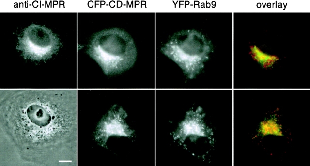
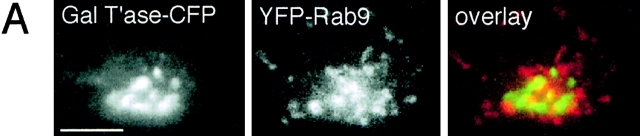
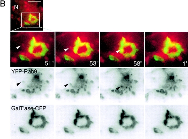
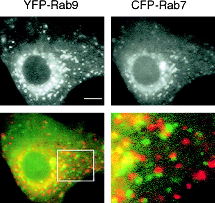
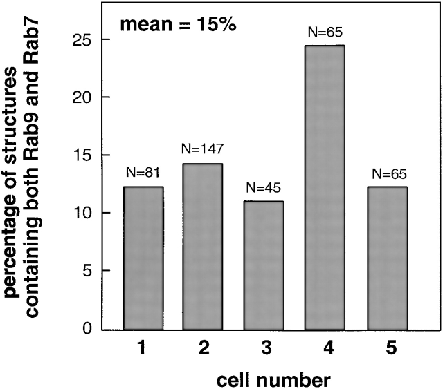
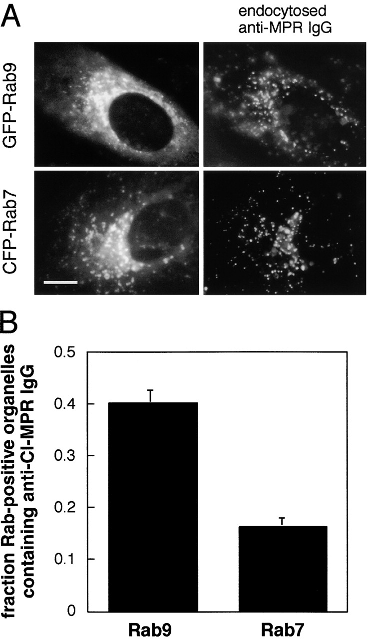
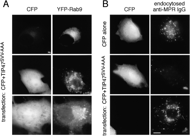
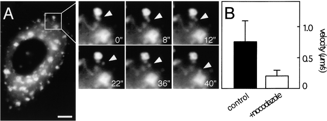
Mannose 6-phosphate receptors (MPRs) are transported from endosomes to the trans-Golgi via a transport process that requires the Rab9 GTPase and the cargo adaptor TIP47. We have generated green fluorescent protein variants of Rab9 and determined their localization in cultured cells. Rab9 is localized primarily in late endosomes and is readily distinguished from the trans-Golgi marker galactosyltransferase. Coexpression of fluorescent Rab9 and Rab7 revealed that these two late endosome Rabs occupy distinct domains within late endosome membranes. Cation-independent mannose 6-phosphate receptors are enriched in the Rab9 domain relative to the Rab7 domain. TIP47 is likely to be present in this domain because it colocalizes with the receptors in fixed cells, and a TIP47 mutant disrupted endosome morphology and sequestered MPRs intracellularly. Rab9 is present on endosomes that display bidirectional microtubule-dependent motility. Rab9-positive transport vesicles fuse with the trans-Golgi network as followed by video microscopy of live cells. These data provide the first indication that Rab9-mediated endosome to trans-Golgi transport can use a vesicle (rather than a tubular) intermediate. Our data suggest that Rab9 remains vesicle associated until docking with the Golgi complex and is rapidly removed concomitant with or just after membrane fusion.


Figures

Figure 1.
Figure 1.
Localization of fluorescent Rab9protein in living cells. BS-C-1 cells expressing CFP-CD-MPR and YFP-Rab9 (second, third, and fourth columns) were analyzed by fluorescence microscopy in comparison with untransfected fixed cells (first column). Endogenous CI-MPRs were detected using mouse anti–CI-MPR culture supernatant. The overlays at right show CFP-CD-MPR (green), YFP-Rab9 (red), and colocalization of both proteins (yellow). Bar, 10 μm.
Figure 8.
Figure 8.
Visualization of a Rab9-bearing vesicle fusing with the trans-Golgi detected by time-lapse video microscopy. See also video 4 available at http://www.jcb.org/cgi/content/full/jcb.200109030/DC1. BS-C-1 cells transfected with pECFP-galactosyltransferase (GalT'ase) and pEYFP-Rab9 were analyzed by fluorescence microscopy. (A) GalT'ase-CFP (left) and YFP-Rab9 (middle) in transfected cells. Overlay of GalT'ase-CFP (green) and YFP-Rab9 (red) is shown on the right. (B) Enlarged selected video images from a time-lapse series of BS-C-1 cells expressing GalT'ase-CFP and YFP-Rab9 proteins. Overlay images (top row) shows GalT'ase-CFP (green), YFP-Rab9 (red), and colocalization at docking and possibly fusion (yellow). Overlay images were obtained by overlapping GalT'ase-CFP signal (middle row) and YFP-Rab9 signal (bottom row). N, nucleus. Bars, 10 μm.
Figure 8.
Figure 8.
Visualization of a Rab9-bearing vesicle fusing with the trans-Golgi detected by time-lapse video microscopy. See also video 4 available at http://www.jcb.org/cgi/content/full/jcb.200109030/DC1. BS-C-1 cells transfected with pECFP-galactosyltransferase (GalT'ase) and pEYFP-Rab9 were analyzed by fluorescence microscopy. (A) GalT'ase-CFP (left) and YFP-Rab9 (middle) in transfected cells. Overlay of GalT'ase-CFP (green) and YFP-Rab9 (red) is shown on the right. (B) Enlarged selected video images from a time-lapse series of BS-C-1 cells expressing GalT'ase-CFP and YFP-Rab9 proteins. Overlay images (top row) shows GalT'ase-CFP (green), YFP-Rab9 (red), and colocalization at docking and possibly fusion (yellow). Overlay images were obtained by overlapping GalT'ase-CFP signal (middle row) and YFP-Rab9 signal (bottom row). N, nucleus. Bars, 10 μm.
Figure 2.
Figure 2.
GFP-Rab9 distribution in BS-C-1 cell extracts subjected to sucrose density gradient flotation. Fractions were collected from the top (fraction 1) to the bottom (fraction 18) of the gradient. Aliquots of each fraction were analyzed by immunoblot using anti-p115 (top) or anti-Rab9 culture supernatant (middle and bottom).
Figure 3.
Figure 3.
YFP-Rab9 and CFP-Rab7 localize to distinct domains on late endosomes. See also video 1 available at http://www.jcb.org/cgi/content/full/jcb.200109030/DC1. YFP-Rab9 (top left) and CFP-Rab7 (top right) in living transfected BS-C-1 cells. Overlay of both images (bottom) shows CFP-Rab7 (green), YFP-Rab9 (red), and the colocalization of both proteins (yellow). The box in the overlay indicates the boundaries of the enlarged image shown in the bottom right panel. Bar, 10 μm.
Figure 4.
Figure 4.
Quantitation of the overlap between Rab9 and Rab7. Cells were transfected with plasmids encoding YFP-Rab9 and CFP-Rab7. Labeled structures containing Rab9, Rab7, or both were counted at high magnification on a computer screen using Metamorph software. Total number of structures counted (N) is shown for five different cells.
Figure 5.
Figure 5.
Distribution of the CI-MPR in Rab9 and Rab7 endosomal subdomains. Cells expressing either GFP-Rab9 or CFP-Rab7 were incubated with Texas red anti–CI-MPR for 3 h and chased for 15 min at 37°C to label a large fraction of total cellular CI-MPRs as shown in A. The proportion of Rab9- or Rab7-positive structures that contained anti–CI-MPR IgG was quantified using Metamorph software on a large computer screen (B). For Rab9, 370 total structures were counted from 8 cells; 146 of these contained anti-MPR IgG. For Rab7, 1,529 vesicles were counted from 15 cells; 250 of these contained MPR IgG. Standard error of the mean was determined using the values obtained for each individual cell analyzed.
Figure 6.
Figure 6.
A TIP47 mutant disrupts the Rab9 compartment and sequesters MPRs intracellularly. (A) Cells stably expressing YFP-Rab9 were transiently cotransfected with plasmids encoding CFP (to detect transfected cells, left column) and TIP47SVV-AAA. The distribution of Rab9 (right column) was visualized by light microscopy. (B) BS-C-1 cells were transfected with a plasmid encoding either CFP alone (top row) or CFP and TIP47SVV-AAA. Cells were then incubated with Texas red anti–CI-MPR at 37°C as in Fig. 5.
Figure 7.
Figure 7.
GFP-Rab9–bearing vesicles display microtubule-based motility. See also videos 2 and 3 available at http://www.jcb.org/cgi/content/full/jcb.200109030/DC1. BS-C-1 cells transfected with pEGFP-Rab9 were treated with or without 4 μg/ml nocodazole for 1 h at 37°C and analyzed by time-lapse video microscopy at 37°C. (A) GFP-Rab9 localization in BS-C-1 nocodazole-treated cells and enlarged selected video images from a time-lapse series. The white arrowhead points to a GFP-Rab9–bearing vesicle. (B) GFP-Rab9 vesicle velocity in BS-C-1–transfected cells with (+ nocodazole) or without (control). Bar, 5 μm.

References

    1. Bucci, C., P. Thomsen, P. Nicoziani, J. McCarthy, and B. van Deurs. 2000. Rab7: a key to lysosome biogenesis. Mol. Biol. Cell. 11:467–480. - PMC - PubMed
    1. Carroll, K.S., J. Hanna, I. Simon, J. Krise, P. Barbero, and S.R. Pfeffer. 2001. Role of the Rab9 GTPase in facilitating receptor recruitment by TIP47. Science. 292:1373–1377. - PubMed
    1. Diaz, E., and S.R. Pfeffer. 1998. TIP47: a cargo selection device for mannose 6-phosphate receptor trafficking. Cell. 93:433–443. - PubMed
    1. Diaz, E., F. Schimmöller, and S.R. Pfeffer. 1997. A novel Rab9 effector required for transport from endosomes to the TGN. J. Cell Biol. 138:283–290. - PMC - PubMed
    1. Draper, R.K., Y. Goda, F.M. Brodsky, and S.R. Pfeffer. 1990. Anti-clathrin antibodies inhibit endocytosis but not receptor recycling to the trans Golgi network in vitro. Science. 248:1539–1541. - PubMed

MeSH terms